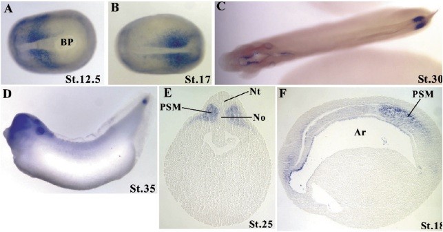
Fig. 3. Spatial expression pattern of Xtbx6r revealed by in situ hybridization. (A-C) Dorsal view. (D) Lateral view. (A-D) Anterior is to the left. (E) Transverse sections, dorsal is to the top. (F) Parasagittal sections, anterior is to the left. Ar, archenteron; Bc, blastocoel; Bp, blastopore; No, notochord; Nt, neural tube; PSM, presomitic mesoderm
Image published in: Yabe S et al. (2006)
Copyright © 2006. Image reproduced with permission of the Publisher.
| Gene | Synonyms | Species | Stage(s) | Tissue |
|---|---|---|---|---|
| tbx6r.L | xtbx6r | X. laevis | Throughout NF stage 12.5 | presumptive paraxial mesoderm |
| tbx6r.L | xtbx6r | X. laevis | Throughout NF stage 17 | paraxial mesoderm posterior |
| tbx6r.L | xtbx6r | X. laevis | Throughout NF stage 18 | paraxial mesoderm |
| tbx6r.L | xtbx6r | X. laevis | Throughout NF stage 25 | presomitic mesoderm |
| tbx6r.L | xtbx6r | X. laevis | Throughout NF stage 29 and 30 | tail tip |
| tbx6r.L | xtbx6r | X. laevis | Throughout NF stage 35 and 36 | tail tip head region otic vesicle |
Image source: Published
Permanent Image Page
Printer Friendly View
XB-IMG-83489