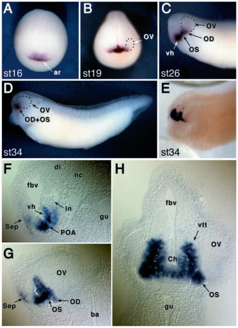
Fig. 6. Developmental expression of XVax1 analyzed by wholemount in situ hybridization. (A) Anterior view of an embryo at stage 16. Staining is detected in the anterior neural ridge. (B) Anterior view of XVax1 expression at stage 19. XVax1 signal is visible in the ventral forebrain. The optic vesicle (OV) shows no expression. (C) Ventral/anterior view of an embryo at stage 26. XVax1 transcripts are detected in the ventral hypothalamus (vh), optic stalk (OS) and the optic disc (OD). (D) Lateral view at stage 34. XVax1 transcripts are located the ventral and anterior forebrain. (E) To improve resolution, a cleared embryo at stage 34 is shown in a dorsal/lateral view. Strong XVax1 expression is visible in the ventral anterior forebrain with a sharp dorsal boarder. (F-H) Sections of stained plastic embedded embryos at stage 34. F; In a sagittal section XVax1 transcripts are detected in the chiasmatic ridge and the ventral hypothalamus. (G) Parasagittal section showing XVax1 expression in the eye disc and in the optic stalk. (H) Transverse section at the level of the optic chiasm. ar, anterior ridge; ba, branchial arch; Ch, chiasmatic ridge; di, diencephalon; fbv, forebrain vesicle; gu, gut; i, infundibulum; nc, notochord; OD, optic disk; OS, optic stalk; OV, optic vesicle; sep, septal/striatal region; vh, ventral hypothalamus; vtt, ventral tegmental tract.
Image published in: Hallonet M et al. (1998)
Copyright © 1998. Image reproduced with permission of the Publisher and the copyright holder. This is an Open Access article distributed under the terms of the Creative Commons Attribution License.
| Gene | Synonyms | Species | Stage(s) | Tissue |
|---|---|---|---|---|
| vax1.S | vax, vax1-a, vax1-b, vax1b, vax1b-a, xvax1, xvax1b | X. laevis | Throughout NF stage 16 to NF stage 19 | anterior neural ridge |
| vax1.S | vax, vax1-a, vax1-b, vax1b, vax1b-a, xvax1, xvax1b | X. laevis | Throughout NF stage 26 | optic nerve hypothalamus optic stalk |
| vax1.S | vax, vax1-a, vax1-b, vax1b, vax1b-a, xvax1, xvax1b | X. laevis | Sometime during NF stage 33 and 34 to NF stage 66 | optic stalk optic nerve ventral tegmentum hypothalamus optic chiasm preoptic area |
Image source: Published
Permanent Image Page
Printer Friendly View
XB-IMG-86874