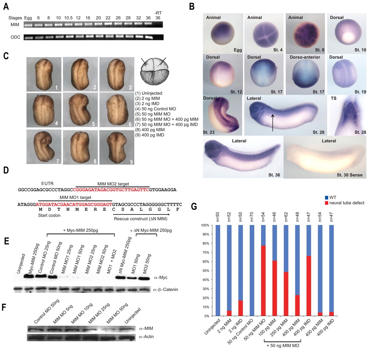
Fig. 4. Temporal and spatial expression pattern of MIM and regulation of anterior neural fold closure by MIM. (A) Xenopus MIM (XMIM) is expressed throughout development as monitored by RT-PCR analysis. ODC is used as a loading control; âRT, without reverse transcriptase. (B) The spatial expression pattern of XMIM is dynamic, with highest expression observed in the neural folds of the neurula stage embryo. Stages and views of the embryos are indicated. No signal is detected using a XMIM sense probe. Arrow indicates the plane of the transverse section (TS). (C) Injection of MIM or IMD RNA produces no morphological defects in the Xenopus embryo, whereas injection of MIM MO (50 ng) dorsally inhibits anterior neural fold closure. The MIM MO phenotype can be effectively rescued by co-injection of 400 pg δN MIM RNA. (D) Location of the MIM MO target sequences; these are absent from the MIM rescue construct (δN MIM), which only includes sequence downstream of the MO1 target. (E) Injection of MIM MO1 or MO2 inhibits translation of Myc-MIM but not of endogenous β-catenin. (F) Injection of MIM MO1 inhibits translation of endogenous MIM but not of endogenous actin in the Xenopus embryo. (G) Quantification of the results of C. The number of injected embryos analyzed is shown above each bar.
Image published in: Liu W et al. (2011)
Copyright © 2011. Image reproduced with permission of the Publisher and the copyright holder. This is an Open Access article distributed under the terms of the Creative Commons Attribution License.
| Gene | Synonyms | Species | Stage(s) | Tissue |
|---|---|---|---|---|
| mtss1.2.L | MIM, mtss1, XMIM | X. laevis | Throughout NF stage 10 to NF stage 12 | dorsal ectoderm neuroectoderm |
| mtss1.2.L | MIM, mtss1, XMIM | X. laevis | Throughout NF stage 17 | anterior neural fold neural plate |
| mtss1.2.L | MIM, mtss1, XMIM | X. laevis | Throughout NF stage 19 | neural plate pre-chordal neural plate chordal neural plate cranial neural crest |
| mtss1.2.L | MIM, mtss1, XMIM | X. laevis | Throughout NF stage 23 | neural tube somite anterior neural fold paraxial mesoderm presomitic mesoderm |
| mtss1.2.L | MIM, mtss1, XMIM | X. laevis | Throughout NF stage 28 to NF stage 35 and 36 | eye retina pharyngeal arch otic vesicle spinal cord somite brain head |
| mtss1.2.L | MIM, mtss1, XMIM | X. laevis | Throughout NF stage 4 (8-cell) | animal animal blastomere |
| mtss1.2.L | MIM, mtss1, XMIM | X. laevis | Throughout NF stage 8 | animal hemisphere |
| mtss1.2.L | MIM, mtss1, XMIM | X. laevis | Throughout unfertilized egg stage | animal hemisphere |
Image source: Published
| Experiment + Assay | Source | Phenotypes and Disease | ||
|---|---|---|---|---|
| Xla Wt + mtss1.2 MO + NF23 (whole-mount microscopy) | Fig 4. C5 |
|
Permanent Image Page
Printer Friendly View
XB-IMG-87095