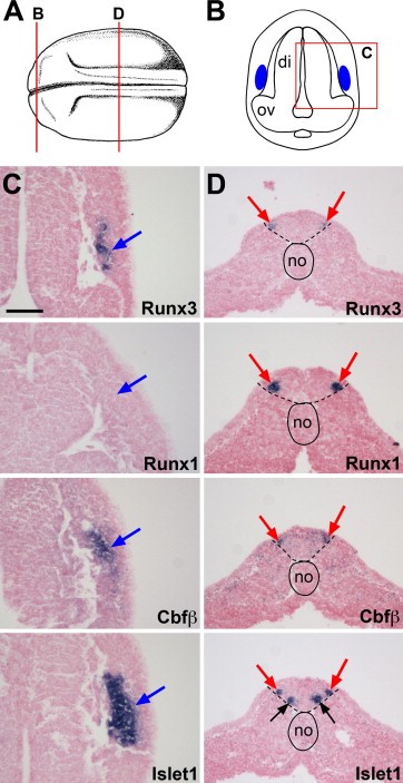
Fig. 5. Comparative analysis of Runx3, Runx1, Cbfβ and Islet1 expression in the profundal-trigeminal placode and Rohon-Beard sensory neurons at stage 20. (A) Schematic representation of a stage 20 embryo view from the dorsal side, anterior to the left. Modified from Nieuwkoop and Faber (1967). (B) Schematic representation of a transverse section at the level of the diencephalon, corresponding to the line labeled âBâ in panel (A). Dorsal to top. The blue areas indicate the position of the developing profundal-trigeminal placode. The boxed area labeled âCâ is the region of the section shown in subsequent panels (C). (C) Runx3, Cbfβ and Islet1 are co-expressed in the profundal-trigeminal placode (blue arrows), while Runx1 is not detected in this tissue. (D) Transverse sections in the trunk region corresponding to the line labeled âDâ in panel (A). All four genes are co-expressed in Rohon-Beard sensory neurons (red arrows). Islet1 is also expressed in the population of ventral interneurons (black arrows). The notochord is outlined with a solid line, while the position of the neural tube is underlined with dashed lines. The probes are indicated in the lower right corner of each panel. di, diencephalon; no, notochord; ov, optic vesicle. The scale bar in panel C represents 100 μm.
Image published in: Park BY and Saint-Jeannet JP (2010)
Copyright © 2010. Image reproduced with permission of the Publisher, Elsevier B. V.
| Gene | Synonyms | Species | Stage(s) | Tissue |
|---|---|---|---|---|
| runx3.L | aml2, cbfa3, pebp2ac | X. laevis | Throughout NF stage 20 | trigeminal placode Rohon-Beard neuron profundal placode neuroectoderm neural plate neural tube |
| runx1.L | aml, aml-1, aml1, aml1-evi-1, aml1 oncogene, amlcr1, cbfa2, evi-1, LOC108709238, pebp2ab, Runx-1, Xaml, Xaml1 | X. laevis | Throughout NF stage 20 | Rohon-Beard neuron neuroectoderm neural tube neuron |
| cbfb.S | cbf-beta, cbfbeta, pea2-beta, pebp2b, pebp2-beta | X. laevis | Throughout NF stage 20 | trigeminal placode Rohon-Beard neuron neural plate profundal placode neuroectoderm neural tube neuron |
| isl1.S | islet-1, islet1, Xisl-1, Xislet-1 | X. laevis | Throughout NF stage 20 | trigeminal placode Rohon-Beard neuron neuron neural tube profundal placode neural plate neuroectoderm |
Image source: Published
Permanent Image Page
Printer Friendly View
XB-IMG-42635