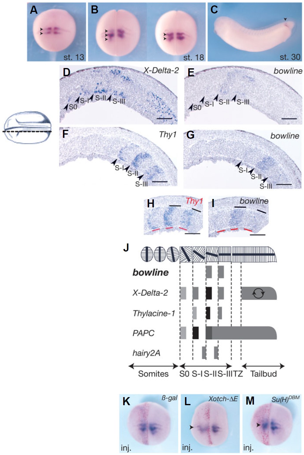
Fig. 2. Localization and transcriptional regulation of bowline transcripts. (A-C) Xenopus embryos from different stages were stained for expression of bowline mRNA by whole-mount in situ hybridization at (A) stage 13, (B) stage 18 and (C) stage 30. Embryos are oriented in dorsal view with anterior to the top except for (C), for which the anterior is to the left. (D-I) Longitudinal section through stage 20 embryos stained for expression of XDelta- 2 (D), bowline (E,G,I) and Thy1 (F,H). Embryos are oriented with anterior to the left. Arrowheads on the lower side of the embryos indicate the anterior end of X-Delta-2-and Thy1-expressing regions. The probes used are shown in the upper right corner. In (H and I), the width of the expression region for Thy1 and bowline are indicated as red and black lines, respectively. (J) Diagram showing the position of bowline expression relative to that of other genes known to be expressed segmentally in X. laevis embryos (Jen et al., 1997, Kim et al., 2000, Sparrow et al., 1998). (KM) Embryos were unilaterally injected with β-gal RNA (K), XotchδE RNA (L), or XSu(H)DBM RNA (M) and analyzed for the expression of bowline (K,L,M) by whole-mount in situ hybridization at stage 20. RNA encoding the lineage tracer β-gal was co-injected to identify the injected side (red staining). Dorsal views with anterior towards the top are shown. Injected sides are indicated as �inj.� Somitomeres were demarcated as S0, S-I, S-II, S-III based on the annotation of Pourqui� and Tam (Pourqui� and Tam, 2001). Bars, 100 μm.
Image published in: Kondow A et al. (2006)
Copyright © 2006. Reproduced with permission of the Publisher, University of the Basque Country Press.
| Gene | Synonyms | Species | Stage(s) | Tissue |
|---|---|---|---|---|
| ripply2.2.S | bowline, LOC100487498, ripply2.2-a, ripply2.2-b | X. laevis | Throughout NF stage 13 | mesoderm presomitic mesoderm paraxial mesoderm |
| ripply2.2.S | bowline, LOC100487498, ripply2.2-a, ripply2.2-b | X. laevis | Throughout NF stage 18 | mesoderm presomitic mesoderm |
| ripply2.2.S | bowline, LOC100487498, ripply2.2-a, ripply2.2-b | X. laevis | Throughout NF stage 29 and 30 | presomitic mesoderm |
| ripply2.2.S | bowline, LOC100487498, ripply2.2-a, ripply2.2-b | X. laevis | Throughout NF stage 20 | presomitic mesoderm |
| dll4.L | delta 2, LOC108705808 | X. laevis | Throughout NF stage 20 | presomitic mesoderm tail bud |
| mespa.S | hen2, mespa-a, mespa-b, thy2, thyl1, thyl2, thylacine 1, thylacine 2, thylacine1, thylacine2 | X. laevis | Throughout NF stage 20 | presomitic mesoderm |
Image source: Published
Permanent Image Page
Printer Friendly View
XB-IMG-83135