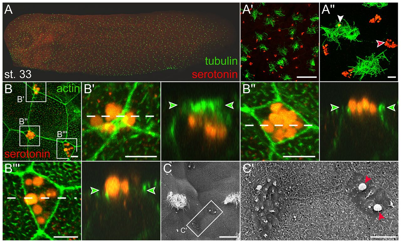
Fig. 1. Small secretory cells (SSCs) in the Xenopus tadpole epidermis. (A) Stage 33 tadpole stained for serotonin (red) and cilia (green) using anti-serotonin and anti-acetylated-α-tubulin antibodies. (Aâ²,Aâ²) Higher magnification reveals vesicular nature of serotonin staining. Please note a serotonin-positive vesicle attached to cilia (white arrowhead in Aâ²). (B) Localization of serotonin-containing vesicles in SSCs. Cell boundaries marked by actin staining using phalloidin. Higher magnifications (left panels in Bâ²-Bâ²â²) and orthogonal projection of z-stack slices (white dashed lines; right panels in Bâ²-Bâ²â²) demonstrate serotonin-positive vesicles beneath, at, or above the apical cell membrane (Bâ²-Bâ²â², green arrowheads). (C) SEM of stage 32 tadpole displaying MCCs and SSCs. (Câ²) Higher magnification of two SSCs. Red arrowheads highlight detaching vesicles. Scale bars: 50 μm in Aâ²; 10 μm in Aâ²; 5 μm in B-Bâ²â²; 20 μm in C; 5 μm in Câ².
Image published in: Walentek P et al. (2014)
Copyright © 2014. Image reproduced with permission of the Publisher.
| Gene | Synonyms | Species | Stage(s) | Tissue |
|---|---|---|---|---|
| tuba4a.L | alpha tubulin, alpha-tubulin, tuba4, tuba4b | X. laevis | Throughout NF stage 33 and 34 | epidermis epithelium cilium secretory epithelial cell |
Image source: Published
Permanent Image Page
Printer Friendly View
XB-IMG-133741