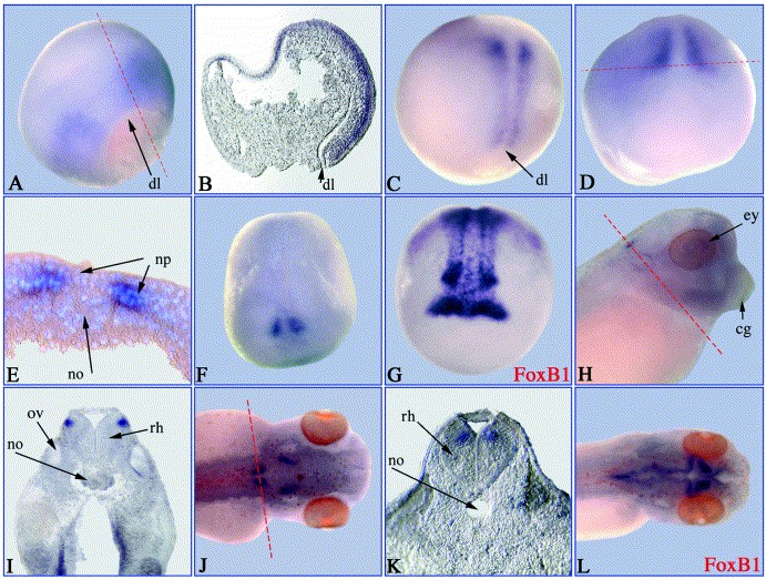
Fig. 3. Spatial expression of xFoxB2. Whole mount in situ hybridisation was performed with xFoxB2 and xFoxB1 (G, L) antisense probes and embryos at different developmental stages. (A) Stage 11; (B) parasagittal section of A; (C) stage 12; (D) stage 14; (E) transversal section of D, nuclei stained with DAPI; (F) stage 15; (G) FoxB1 (Fkh-5), stage 16; (H) stage 35; (I) horizontal section of H; (J) stage 41; (K) horizontal section of J; (L) FoxB1, stage 41. Embryos shown in (AâC) are orientated with dorsal side to the right and animal half to the top; D, F, G anterior view; H shows a lateral and J as well as L a dorsal view. The plane of sections is marked by red lines. cg, cement gland; dl, dorsal lip; ey, eye; np, neural plate; no, notochord; ov, otic vesicle; rh, rhombencephalon.
Image published in: Pohl BS et al. (2002)
Copyright © 2002. Image reproduced with permission of the Publisher, Elsevier B. V.
| Gene | Synonyms | Species | Stage(s) | Tissue |
|---|---|---|---|---|
| foxb2.S | LOC108696911, xfd-5, xFoxB2 | X. laevis | Throughout NF stage 11 | upper blastopore lip |
| foxb2.S | LOC108696911, xfd-5, xFoxB2 | X. laevis | Throughout NF stage 14 | notochord neural plate |
| foxb2.S | LOC108696911, xfd-5, xFoxB2 | X. laevis | Throughout NF stage 35 and 36 | cement gland eye notochord otic vesicle hindbrain |
| foxb2.S | LOC108696911, xfd-5, xFoxB2 | X. laevis | Throughout NF stage 41 | notochord hindbrain |
Image source: Published
Permanent Image Page
Printer Friendly View
XB-IMG-45408