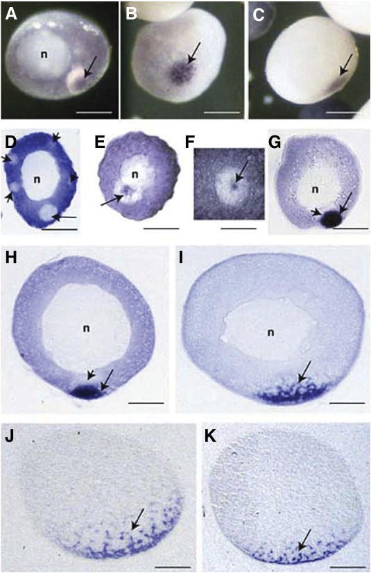
Fig. 3. Centroid mRNA localization in Xenopus oocytes. (A-C) Whole mount in situ hybridization showing localization of centroid mRNA (arrows) in the mitochondrial cloud in a stage 1 oocyte (A) and in the apex of the vegetal cortex in a stage II oocyte (B) and a stage III oocyte (C). (D- J) Sections of whole mount in situ hybridization showing localization of centroid mRNA in oocytes at different stages. (D) In a pre-stage I oocyte, centroid mRNA is uniformly dispersed in the cytoplasm but excluded from the mitochondrial cloud (long arrow) and secondary clouds (short arrows). (E,F) In a stage I oocyte, centroid mRNA (arrow) is located in the center of the mitochondrial cloud. Panel (F) shows the high magnification of the mitochondrial cloud (white sphere) with centrally located centroid mRNA (arrow). (G,H) In late stage I/early stage II and stage II oocytes, centroid mRNA is limited to the vegetal tip of the mitochondrial cloud (co- localizing with the germ plasm [long arrow]) and is excluded from the apical part of the mitochondrial cloud (short arrows). (I-K) Stage III, early stage IV and stage IV oocytes showing localization of centroid mRNA in the islands of a dispersing mitochondrial cloud (arrows). n, nucleus. Scale barsareequalto56μmin(A),90μmin(B,G),100μmin(C),70μmin(D), 75 μm in (E), 65 μm in (F), 86 μm in (H), 80 μm in (I) and 100 μm in (J,K).
Image published in: Kloc M and Chan AP (2007)
Copyright © 2007. Image reproduced with permission of the Publisher, University of the Basque Country Press.
| Gene | Synonyms | Species | Stage(s) | Tissue |
|---|---|---|---|---|
| ddx59.L | centroid | X. laevis | Sometime during oocyte stage II to oocyte stage III | mitochondrial cloud |
| ddx59.L | centroid | X. laevis | Sometime during oocyte stage IV to oocyte stage V | vegetal cortex mitochondrial cloud |
| ddx59.L | centroid | X. laevis | Throughout unfertilized egg stage | cytoplasm |
Image source: Published
Permanent Image Page
Printer Friendly View
XB-IMG-76290