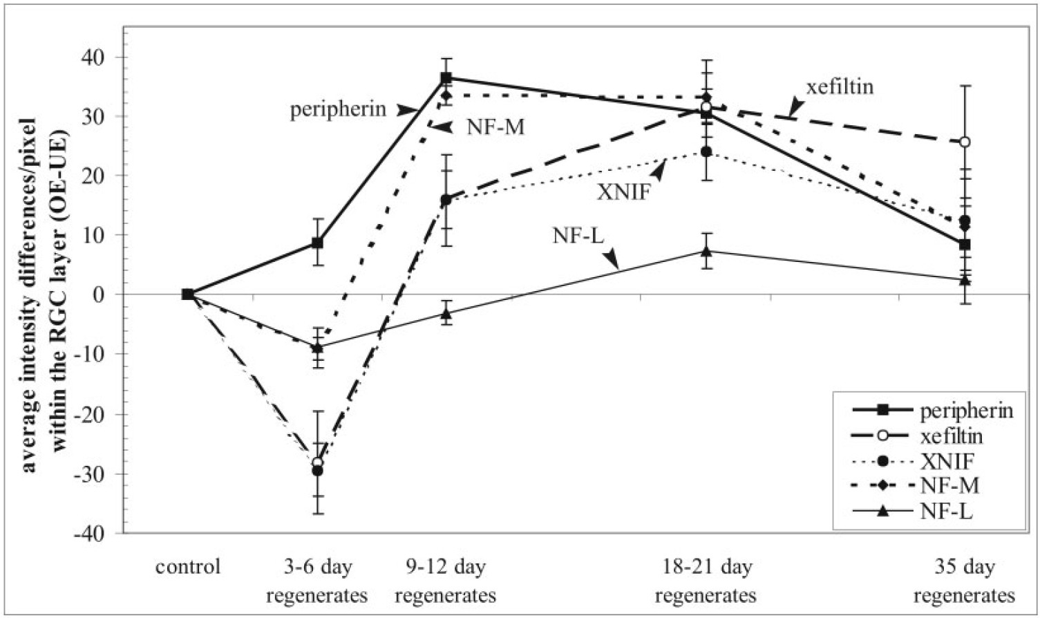
Fig. 4. Differences in nIF mRNA levels between the operated (OE) and unoperated, contralateral control (UE) RGCs, as measured by video densitometry from in situ hybridizations performed under standardized conditions. For each nIF and time period after optic nerve crush, the difference in the mean intensity/pixel was determined between the UE and OE RGCs and then averaged among animals from a given time period (mean SE; n 4 animals for 3â6 days and 9â12 days; 5 for 18â21 days; 2 for 35 days). Levels of nIF mRNA remain relatively stable from 3â6 days postcrush, when regenerating axons cross the lesion and enter the optic nerve, from 9â12 days, when they have reached the optic chiasm, and from 18â21 days, when they reach and cover the tectum. Data within each of these time windows were therefore pooled.
Image published in: Gervasi C et al. (2003)
Copyright © 2003. Image reproduced with permission of the Publisher, John Wiley & Sons.
Permanent Image Page
Printer Friendly View
XB-IMG-86776