XB-IMG-171571
Xenbase Image ID: 171571
|
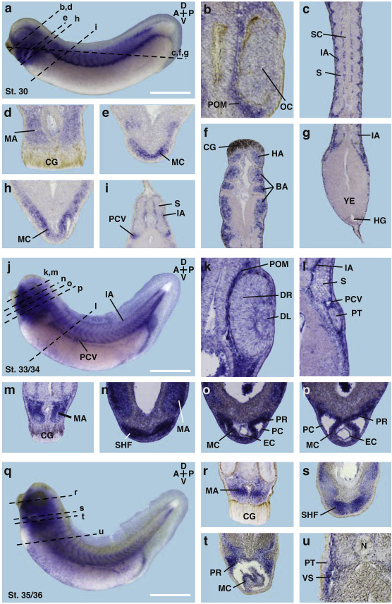
Fig. 6. Sections of adgra2 stained Xenopus late tadpoles.
Spatial expression pattern of adgra2 is visualized by WMISH followed by cross sectioning of Xenopus embryos. a/j/q. Lateral views with anterior to the left at indicated stages. Dashed lines indicate the level of sections. b-g. Transversal (b, d, e, h, i) and horizontal (c, f, g) vibratome sections at stage 30. b. adgra2 is present in the periocular mesenchyme (POM). c. adgra2 transcripts are localized in the intersomitic arteries (IA). d. adgra2 is visualized in the mandibular arches (MA). e. adgra2 is expressed in the myocardium (MC) of the developing heart. f. adgra2 expression in the hyoidal (HA) and branchial arches (BA). g. Caudal section with adgra2 transcripts in the intersomitic arteries (IA). h. adgra2 is expressed in the myocardium (MC). i. adgra2 is detected in the posterior cardinal vein (PCV) and the intersomitic arteries (IA) surrounding the somites (S). k-p. Transversal vibratome sections at stage 33/34. k. adgra2 expression is detected in the periocular mesenchyme (POM). l. adgra2 expression in the intersomitic arteries (IA) and the endothelium of the posterior cardinal vein (PCV). m. adgra2 is present in the mandibular arch (MA). n-p. Transcripts of adgra2 are visualized in the heart including the second heart field (SHF), the myocardium (MC), the pericardial roof (PR), the endocard (EC) and the pericard (PC). r-u. Transversal sections at stage 35/36. r. adgra2 is visualized in the mandibular arch (MA). s-t. adgra2 expression is visible in the second heart field (SHF), myocardium (MC), the pericardial roof (PR), and the pericard (PC). r-u. adgra2 transcripts are found in the vascular system (VS) nearby the pronephric tubule (PT). A, anterior; BA, branchial arch; CG, cement gland; D, dorsal; DL, developing lens; DR, developing retina; EC, endocard; HA, hyoidal arch; HG, hindgut; IA, intersomitic arteries; MA, mandibular arch; MC, myocardium; OC, optic cup; P, posterior; PC, pericardium; PCV, posterior cardinal vein; POM, periocular mesenchyme; PR, pericardial roof; PT, pronephric tubule; S, somite; SC, spinal cord; SHF, Second heart field; V, ventral; VS, vascular system; YE, yolk endoderm. Scale bars: 1â¯mm (a, j, q). Image published in: Seigfried FA et al. (2018) Copyright © 2018. Image reproduced with permission of the Publisher, Elsevier B. V.
Image source: Published Larger Image Printer Friendly View |