XB-IMG-123197
Xenbase Image ID: 123197
|
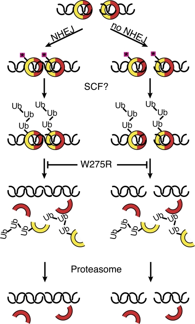
Figure 8. A model for Ku removal from DNA. Ku70 (red) and Ku80 (yellow) form a ring that encircles a DSB. The heterodimer either recruits other NHEJ proteins, repairing the break (left), or fails to do so (right). Binding to DNA causes Ku to become posttranslationally modified, possibly through phosphorylation (pink boxes). An E3 ubiquitin ligase such as the SCF complex recognizes this modification and polyubiquitylates Ku80. This leads to dissociation from DNA, which is inhibited by the W275R mutation. Dissociation is followed by proteasomal degradation. Image published in: Postow L et al. (2008) © 2008 Postow et al. Creative Commons Attribution-NonCommercial-ShareAlike license Larger Image Printer Friendly View |