XB-IMG-121551
Xenbase Image ID: 121551
|
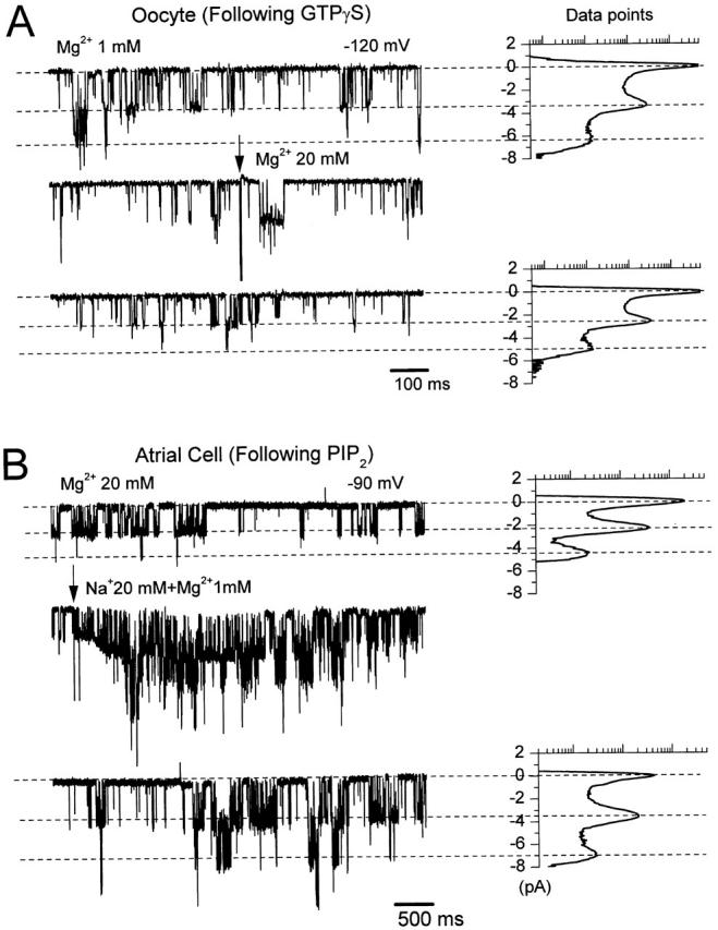
Figure 6. High concentrations of Mg2+ ions reduce the GIRK single-channel currents. (A) Single-channel records of GIRK channels in an inside-out patch excised from an oocyte expressing GIRK1/GIRK4. The membrane was clamped at −120 mV and 5 μM acetylcholine was present in the pipette. The channel was preactivated by 10 μM GTPγS and the current traces shown were recorded after washout of the GTP analogue. The switch between bath solutions containing 1 and 20 mM Mg2+ is visualized by the arrow (and by the corresponding electrical artifact on the record) on top of the second current trace. Associated all-point histogram plots indicate the amplitudes resulting from the various activity levels ranging from closed to multiple open levels and are shown for the first (1 mM Mg2+) and third (20 mM Mg2+) current traces. Data points are shown on a logarithmic scale ranging from 5 to 50,000. (B) KACh channel activity in an inside-out patch from a cardiac cell. The membrane was held at −90 mV and the pipette contained 5 μM acetylcholine. The patch was preincubated with 5 μM PIP2 and the current traces shown were recorded after the washout of PIP2. The arrow on top of the second current trace visualizes the switch between bath solutions containing 20 mM Mg2+ and 20 mM Na+ + 1 mM Mg2+. Associated all-point histogram plots are shown for the first (20 mM Mg2+) and third (20 mM Na+ + 1 mM Mg2+) current traces. Data points are shown on a logarithmic scale ranging from 20 to 50,000. Image published in: Petit-Jacques J et al. (1999) © 1999 The Rockefeller University Press. Creative Commons Attribution-NonCommercial-ShareAlike license Larger Image Printer Friendly View |