XB-IMG-128422
Xenbase Image ID: 128422
|
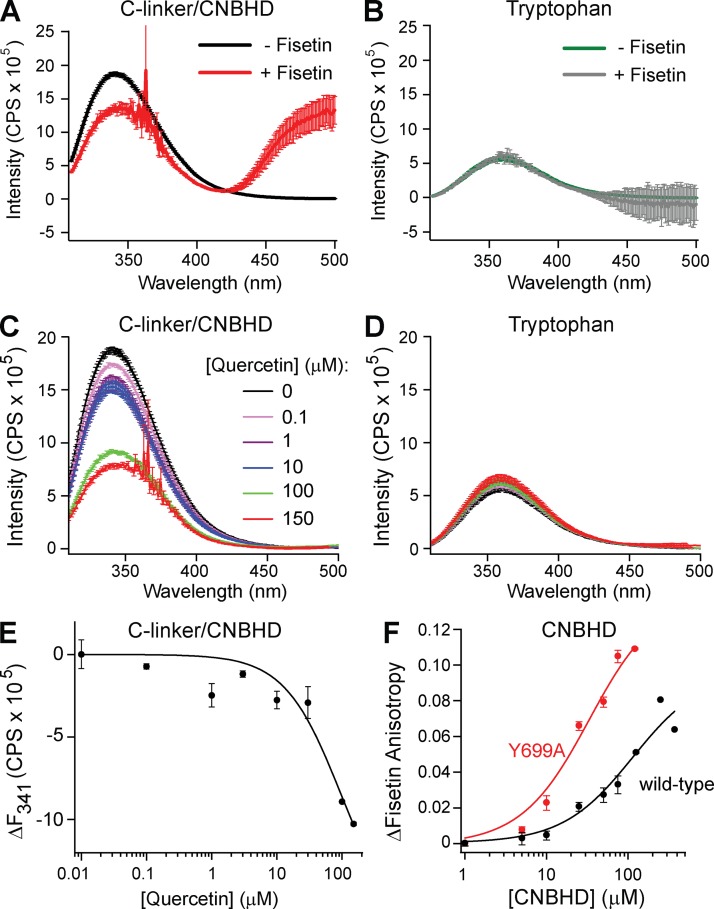
Figure 7. Quercetin and fisetin bound to purified CNBHD of mEAG1 channels. (A and B) The inner filter–corrected and background-subtracted emission spectra of 4 µM of purified C-linker/CNBHD (A) and 4 µM of free tryptophan (B) recorded with 0 or 100 µM fisetin as indicated. (C and D) The inner filter–corrected and background-subtracted tryptophan fluorescence emission spectra of 4 µM of purified C-linker/CNBHD (C) or 4 µM of free tryptophan (D) recorded with various concentrations of quercetin, as indicated. (E) Plot of the change in the peak emission fluorescence intensity (at 341 nm) of the C-linker/CNBHD versus total quercetin concentration. These data were fit with Eq. 6 to report an apparent binding affinity of 93 ± 69 µM. (F) Fluorescence anisotropy of fisetin plotted versus the total concentration of wild-type or Y699A mutant CNBHD. The data were fit with Eq. 7 to yield Kd’s of 111 µM for wild-type and 30 µM for the Y699A mutant. Image published in: Carlson AE et al. (2013) © 2013 Carlson et al. Creative Commons Attribution-NonCommercial-ShareAlike license Larger Image Printer Friendly View |