XB-IMG-158087
Xenbase Image ID: 158087
|
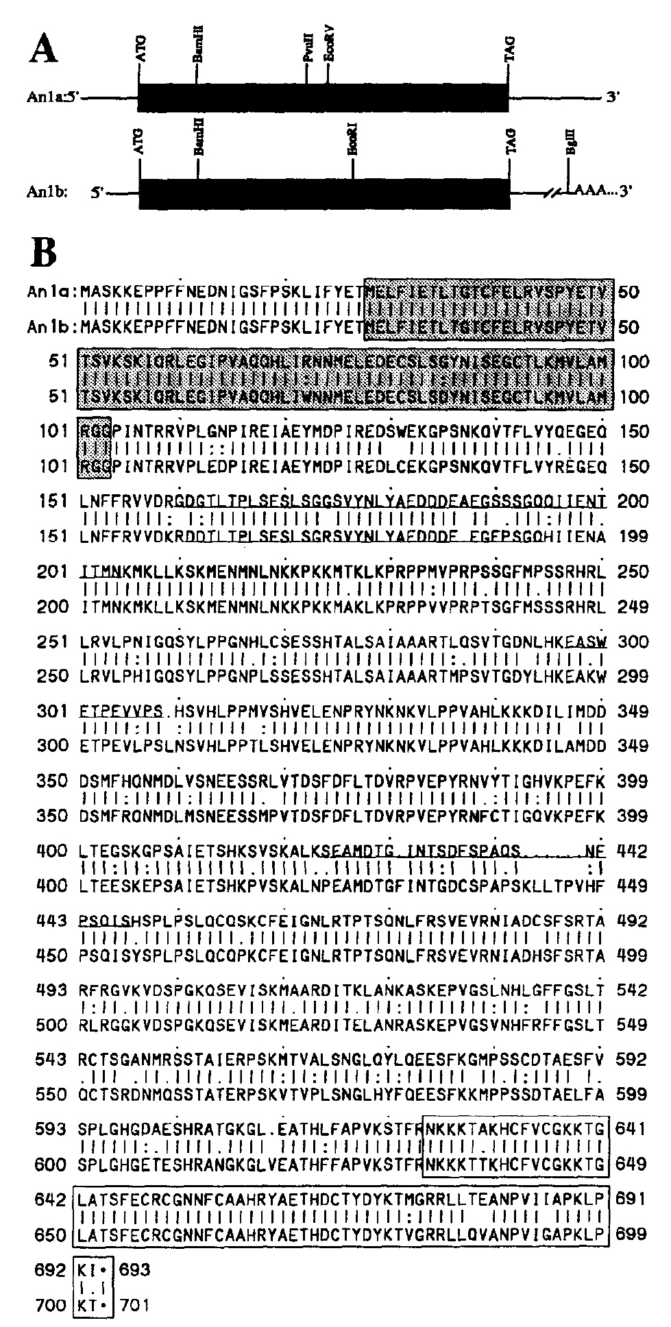
Fig. I. Restriction maps of Anfa and Anlb cDNA clones and alignment
of the two deduced proteins. (A) Restriction maps of Ada and An/b
are shown. The nearly full-length Ada and Anlb cDNAs are approximately
2.9 kb and 2.8 kb and encode proteins predicted to be 76.9 kDa
and 78.5 kDa, respectively. (B) An alignment of the two proteins
encoded by the Ada and Anfb mRNAs according to the algorithm of
Needleman and Wunsch (1970) is shown. Solid vertical lines indicate
exact aa matches; single and double dots indicate conservative replacements.
The shaded box (aa 28-103) shows the region of Ub homology
found in both proteins. The open box (aa 625-693 in An la; aa 633-701
in Anl b) indicates homology to the PvPR3 protein from Phaseolus
vulgaris which includes the putative Zn 2’-finger-metal-binding domain
common to both proteins. Underlined aa are possible PEST sequences
identified using the PEST-FIND program (Rogers et al., 1986). Ada
and Anlb have GenBank accession Nos. LO8474 and L08475. Methods:
cDNA clones were isolated from a phage hgt IO oocyte cDNA library
by screening with the original 650-bp An1 isolate (Rebagliati et al.,
1985). Additional clones were obtained from the same oocyte library by PCR (Friedman et al., 1990) and RT-coupled PCR amplification of
oocyte RNA (Kawasaki, 1990). DNA sequences were obtained for both
strands by the dideoxynucleotide chain-termination procedure (Sanger
et al., 1977) using [a-“S]dATP and the Sequenase” Version 2.0 kit
(Tabor and Richardson, 1987). All nt and aa sequences were analyzed
using the sequence analysis software package of the Genetics Computer
Group, Madison, WI (Devereux et al., 1984). Image published in: Linnen JM et al. (1993) Copyright © 1993. Image reproduced with permission of the Publisher. Larger Image Printer Friendly View |