XB-IMG-124491
Xenbase Image ID: 124491
|
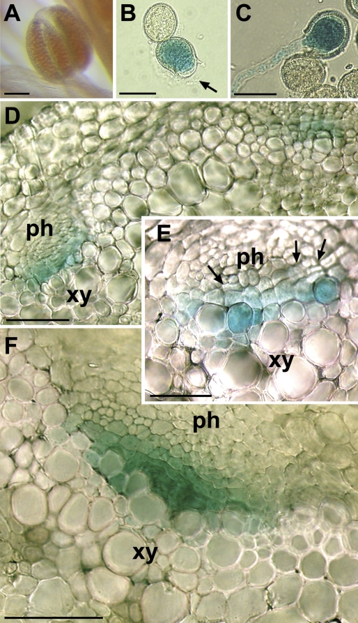
Fig. 6. GUS stainings of pAtPMT1/GUS plants. (A) Anther of a pAtPMT1/GUS plant with no GUS activity. (B) Closed and just opening (arrow) pollen grains on agar medium. (C) Ungerminated (white) and germinated (blue) pollen grains, the latter with a well-developed pollen tube. (D) Cross-section through a flower stalk showing GUS histochemical staining in the centre of two vascular bundles. (E, F) Higher magnifications of cross-sections from flower stalk vascular bundles (ph, phloem; xy, xylem). Arrows show regions, with the typical stacks of cambium and newly formed phloem and xylem vessels. Bars are 100 μm in (A), 20 μm in (B, C), and 25 μm in (D–F). Larger Image Printer Friendly View |