XB-IMG-129523
Xenbase Image ID: 129523
|
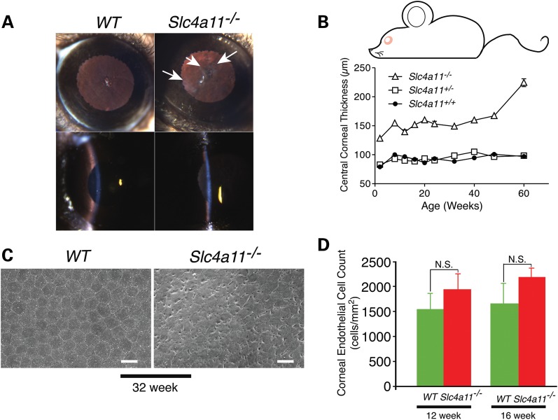
Figure 5. Corneal oedema and progressive corneal dysfunction in slc4A-a11−/− mice. (A) Slit lamp examination of WT and slc4a11−/− mice. Corneas of 48-week WT and slc4a11−/− mice seen under retro-illumination (top panels) and examined using tangential slit illumination (bottom panels). Arrows indicate corneal haze observed in slc4a11−/− mice. (B) Corneal thickness measured by in vivo confocal microscopy (n = 5 per genotype). Central corneal thickness was significantly different between WT and slc4a11−/− mice at all time points (P < 0.01). The corneal thickness in heterozygous (slc4a11+/−) mice was not different compared with wild-type mice. (C) Scanning electron micrographs of corneal endothelia from WT and slc4a11−/−mice at 32 weeks. Scale bars are 20 µm. (D) Statistical analysis of corneal endothelial cell density. Image published in: Vilas GL et al. (2013) © The Author 2013. Creative Commons Attribution license Larger Image Printer Friendly View |