XB-IMG-117183
Xenbase Image ID: 117183
|
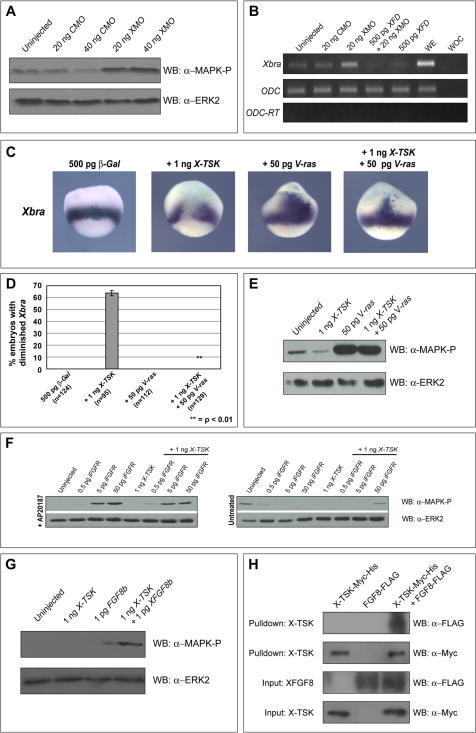
Figure 6. X-TSK inhibition and binding of FGF8b.(A) Western blotting of MAPK phosphorylation in animal caps injected with 20–40 ng CMO and 20–40 ng XMO. Depletion of X-TSK with XMO activates MAPK phosphorylation. (B) Semi-quantitative RT-PCR of Xbra expression in DMZ injected with 20 ng CMO, 20 ng XMO, 500 pg XFD and 500 pg XFD with 20 ng XMO. WE = Whole embryo, WOC = Water only control. Inhibition of FGF signals with XFD blocks Xbra expression activated upon depletion of X-TSK with XMO. (C) Whole mount in situ hybridization of Xbra in stage 10.5 embryos, lateral orientation. Embryos injected with 500 pg β-Gal with 1 ng X-TSK, 50 pg V-ras and X-TSK with V-ras. V-ras blocks X-TSK mediated inhibition of Xbra expression in 100% of embryos analyzed (p = <0.01), represented graphically in (D). (E) Western blotting of MAPK phosphorylation in animal caps injected with X-TSK and V-ras. V-ras blocks X-TSK mediated inhibition of MAPK phosphorylation. (F) Western blotting of MAPK phosphorylation in animal caps injected with X-TSK and iFGFR, in the presence or absence of chemical dimerisation agent, AP20187. Induced dimerisation blocks the activity of X-TSK to inhibit MAPK phosphorylation. (G) Western blotting of MAPK phosphorylation in animal caps injected with X-TSK and FGF8b. X-TSK inhibits MAPK phosphorylation activated by FGF8b. (H) Western blotting of nickel bead pulldown of FGF8b-FLAG in complex with X-TSK-Myc-His. Top panel: detection of FGF8b-FLAG in complex with X-TSK-Myc-His (third lane). Second panel: detection of X-TSK-Myc-His pulled down. Third and bottom panels: detection of FGF8b-FLAG and X-TSK-Myc-His input into the pulldown reaction. Image published in: Morris SA et al. (2007) Morris et al. Creative Commons Attribution license Larger Image Printer Friendly View |