XB-IMG-118989
Xenbase Image ID: 118989
|
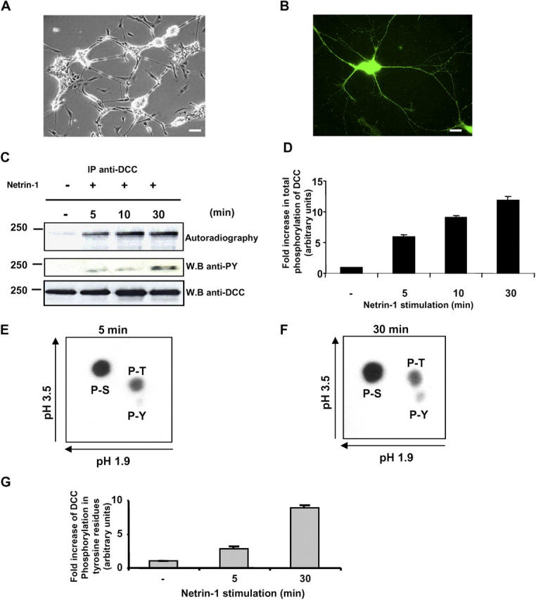
Figure 1. DCC is rapidly phosphorylated in vivo upon Netrin-1 stimulation in embryonic rat CN. (A) Phase-contrast image of E13 rat CN 72 h after plating on laminin. Bar, 40 μm. (B) Immunofluorescence of CN with anti-DCC antibodies showing DCC expression in the cell bodies and along the axons. Bar, 20 μm. (C) E13 rat CN labeled with [32P]orthophosphate for 2 h were either stimulated or not with Netrin-1 for 5, 10, and 30 min. Endogenous DCC was immunoprecipitated (IP) from the cell lysates. The radiolabeled proteins were subjected to SDS-PAGE and identified by autoradiography. The membrane was immunoblotted with anti-pY and anti-DCC antibodies to show the total amount of DCC. (D) Quantitative analysis of the phosphorylation level of DCC after Netrin-1 stimulation of rat CN. Fold increase in total phosphorylation of DCC was determined by densitometry (n = 3). Error bars represent SD. (E and F) The bands corresponding to phosphorylated DCC obtained after 5 min (E) or 30 min (F) of Netrin-1 stimulation were subjected to a phospho–amino acid analysis. (G) Quantitative analysis of DCC phosphorylation on tyrosine residues, using the method described in D (n = 3). Error bars represent SD. Image published in: Meriane M et al. (2004) Copyright © 2004, The Rockefeller University Press. Creative Commons Attribution-NonCommercial-ShareAlike license Larger Image Printer Friendly View |