Click here to close
Hello! We notice that you are using Internet Explorer, which is not supported by Xenbase and may cause the site to display incorrectly.
We suggest using a current version of Chrome,
FireFox, or Safari.
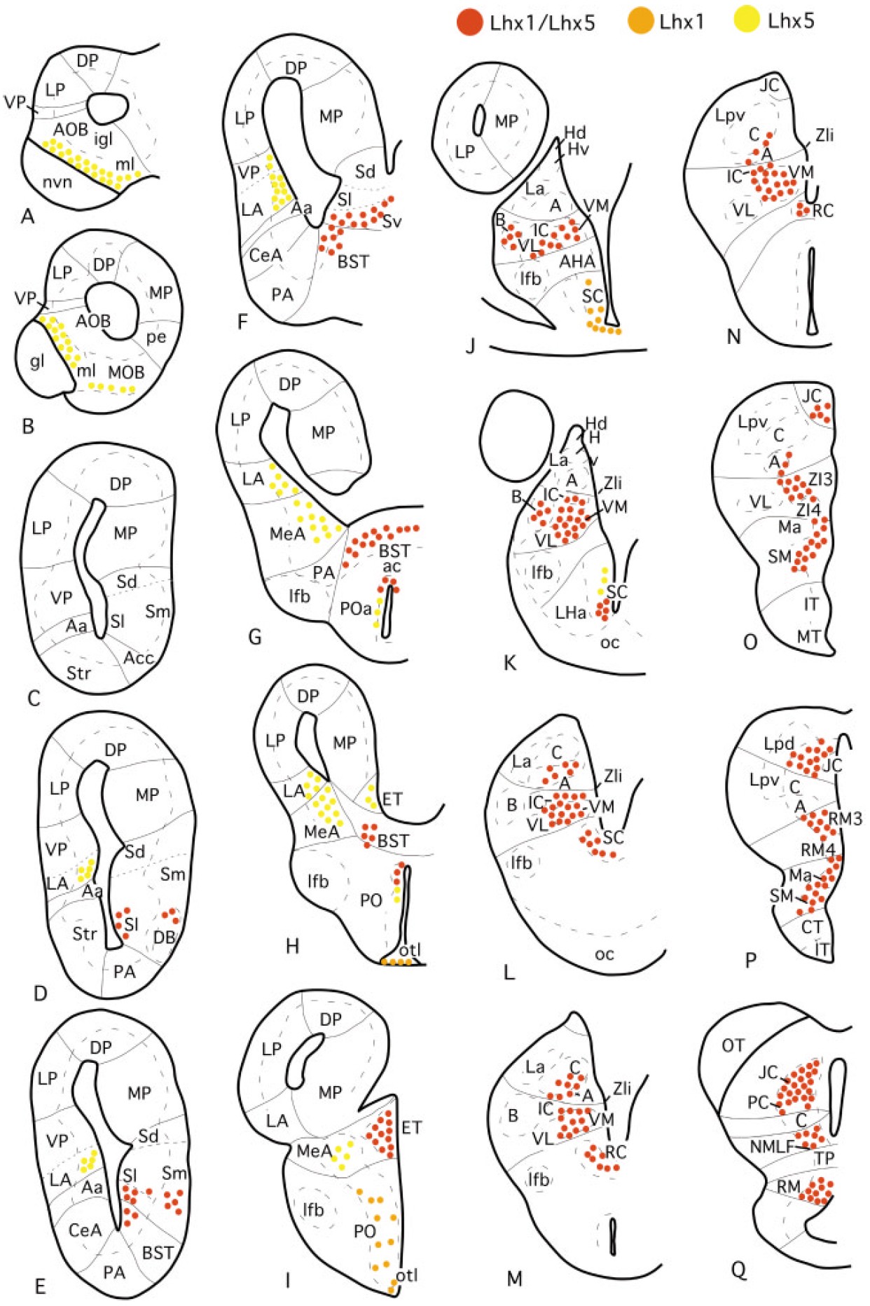
Fig. 2. AâQ: Schematic drawings of transverse sections through the brain of adult Xenopus laevis
illustrating the distribution of x-Lhx1-, x-Lhx5-, and x-Lhx1/5-expressing cells in the forebrain. The
appropriate levels of the sections are indicated in Figure 1. For abbreviations, see list. [Color figure can
be viewed in the online issue, which is available at www.interscience.wiley.com.]
brain forebrain preoptic area suprachiasmatic nucleus telencephalon septum lateral septum medial septum ventral septum bed nucleus of the stria terminalis anterior commissure lateral hypothalamic nucleus ventromedial thalamic nucleus ventrolateral thalamic nucleus central nucleus of the thalamus pretectum mammilary region