XB-IMG-159747
Xenbase Image ID: 159747
|
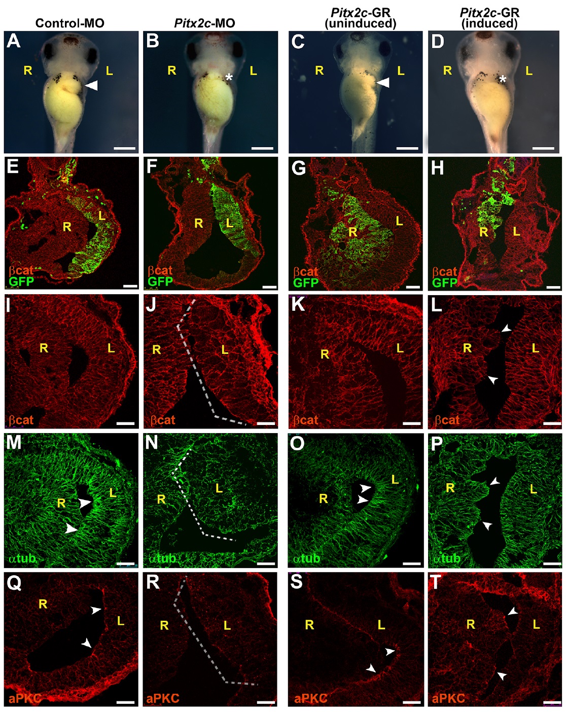
Fig. 4. Pitx2c controls epithelial morphogenesis in left stomach wall. Frog embryos were injected with control
morpholino (control-MO; A,E,I,M,Q) or Pitx2c-MO (B,F,J,N,R) targeted to the left side of the stomach, or injected with Pitx2c- GR mRNA (C,G,K,O,S and D,H,L,P,T) targeted to the right side. (See Fig. S6A,B for morpholino validation.) In injected
embryos (A-D), the greater curvature of the stomach at stage 42 is indicated by an arrowhead (A,C); absence of curvature is
specified by an asterisk (B,D). Sections through stage 39 stomachs (E-T) were stained for β-catenin (βcat; red; E-L),
α-tubulin (αtub; green; M-P) or atypical PKC (aPKC; red; Q-T). GFP mRNA was coinjected as a lineage tracer to confirm proper targeting (green; E-H). MO depletion on the left (F) or ectopic activity of Pitx2 on the right (H) results in a loss of asymmetry
within the stomach compared with controls (E,G, respectively). In addition, compared with control-MO injected embryos, in which αtub and aPKC are concentrated at the apical surface of the left stomach wall (arrowheads in M,Q, respectively), MO
depletion of Pitx2c disrupts epithelial architecture (brackets in J,N,R). Meanwhile, dexamethasone induction of Pitx2c activity in the right wall polarizes stomach endoderm, as indicated by ectopic regions of polarized epithelial architecture (arrowheads in L,P,T), correlating with ectopic αtub (P) and aPKC (T), which are not observed in right wall of uninduced controls (K,O,S). Scale bars: 500 μm in A-D; 75 μm in E-H; 50 μm in I-T. L, left; R, right. Image published in: Davis A et al. (2017) Copyright © 2017. Image reproduced with permission of the Publisher and the copyright holder. This is an Open Access article distributed under the terms of the Creative Commons Attribution License.
Image source: Published
Larger Image Printer Friendly View |