XB-IMG-122880
Xenbase Image ID: 122880
|
|
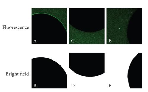
Figure 5. Fluorescence images (top row) and
bright-field images (bottom row) of oocytes incubated with qdot-containing
compounds for 10 minutes. The
bright-field images illustrate the plane of focus of the opaque oocyte. Panels
A and B show results from a human ρ1 GABAC-expressing oocyte incubated with 34 nM muscimol-conjugated AMP-coated qdots. Panels C and D show a human ρ1 GABAC-expressing oocyte incubated with a 34 nM solution of unconjugated AMP-coated qdots. Panels
E and F show a nonexpressing oocyte incubated with 34 nM muscimol-conjugated
AMP-coated qdots. Adapted from Gussin et al. [65]. Image published in: Tomlinson ID et al. (2007) Image downloaded from an Open Access article in PubMed Central. Copyright © 2007 Ian D. Tomlinson et al. Larger Image Printer Friendly View |