XB-IMG-125117
Xenbase Image ID: 125117
|
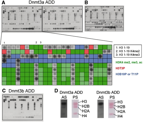
Figure 1. Binding of the Dnmt3a and Dnmt3b ADD domains to peptide arrays and native histones. (A) Binding of Dnmt3a ADD domain to peptide arrays comprising 384 different peptides. The enlargement shows the binding to the H3 1–19 peptides. Peptides containing H3K4me2 or me3, H3T3P, H3S10P or H3T11P are shaded green, red and blue, respectively. The positions of the unmodified H3 1–19 as well as the peptides di- and trimethylated at K4 are annotated. (B) Design of the CelluSpots histone tail peptide arrays. For a detailed annotation of all spots cf. Supplementary Data S1. (C) Binding of Dnmt3b ADD domain to peptide arrays comprising 384 different peptides. (D) Binding of ADD domains to native histones isolated from human cells. Histones were separated by polyacrylamide gel electrophoresis and blotted to Nitrocellulose membrane. The membrane was stained with Ponceau S (PS). Then, membranes were incubated with GST tagged ADD domains, washed and ADD binding detected with anti-GST antibody staining (AS). Image published in: Zhang Y et al. (2010) © The Author(s) 2010. Creative Commons Attribution-NonCommercial license Larger Image Printer Friendly View |