XB-IMG-124866
Xenbase Image ID: 124866
|
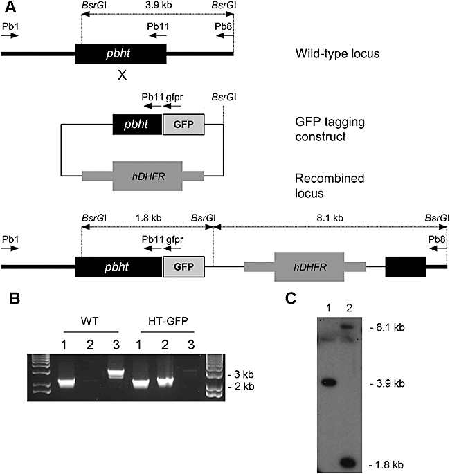
Fig. 5. Tagging of the pbht locus with GFP. A. The strategy for GFP-tagging of the pbht locus; arrows indicate the location of PCR primers. B. PCR analysis of the wild type (WT) and a pyrimethamine-resistant pbht-gfp transfected line. Lane 1, positive control 2.3 kb (primers Pb1 + Pb11); lane 2, detection of the gfp-tagged locus 2.4 kb (Pb1 + gfpr); lane 3, detection of the wild-type locus 3.2 kb (Pb1 + Pb8). C. Southern blot analysis of wild-type (1) and pbht-gfp transfected line (2). gDNA was digested with BsrGI, and blot was probed with a pbht fragment used for generation of the tagging construct; wild-type locus 3.9 kb, integration bands 1.8 kb (5′) and 8.1 kb (3′). Image published in: Slavic K et al. (2010) Journal compilation © 2010 Blackwell Publishing. Creative Commons Attribution license Larger Image Printer Friendly View |