XB-IMG-121119
Xenbase Image ID: 121119
|
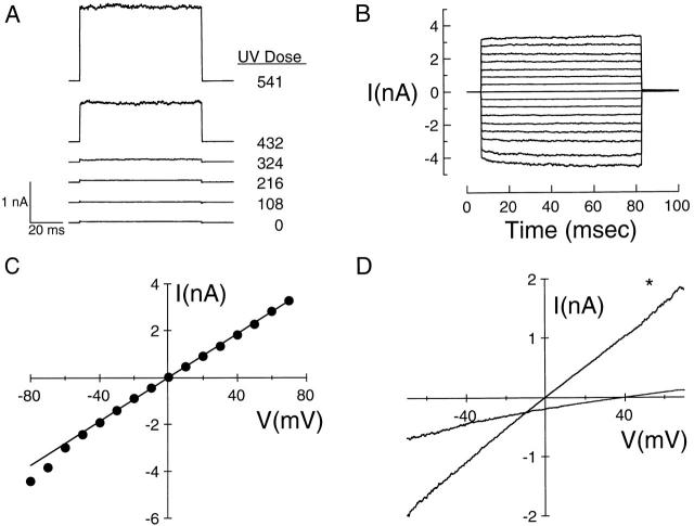
Figure 4. Characterization of UV-activated conductance in Xenopus oocytes. (A) UV effect on the current through an excised, inside-out patch from an uninjected oocyte in response to a 0–50-mV voltage step. UV doses (in photons × 108 · μm−2) are indicated. The patch was irradiated in the absence of cGMP while held at 0 mV. (B) Voltage dependence of UV-activated conductance. Results for patch from A after exposure to 5.41 × 10¹⁰ photons · μm−2. Currents were elicited by stepping the patch from a holding potential of 0 mV to voltages from −80 to +70 mV in 10-mV increments. (C) Current–voltage relation (•) for the results in B. The fit is a straight line with slope 4.6 × 10⁻⁸ S. (D) Cation selectivity of UV-activated conductance. Currents were elicited by voltage ramps from −75 to +75 mV in 500 ms after exposing the patch to 1.78 × 10¹⁰ photons · μm−2. The trace indicated by * was obtained in symmetric 130-mM NaCl solutions; the other trace was obtained with 130 mM NaCl in the pipet and 26 mM NaCl in the bath. Voltages were not corrected for junction potentials. The results are from a different patch than that used in A–C. The currents in A–D were corrected for leak through the seal resistance by subtracting the current before irradiation. Image published in: Middendorf TR et al. (2000) © 2000 The Rockefeller University Press. Creative Commons Attribution-NonCommercial-ShareAlike license Larger Image Printer Friendly View |