XB-IMG-129394
Xenbase Image ID: 129394
|
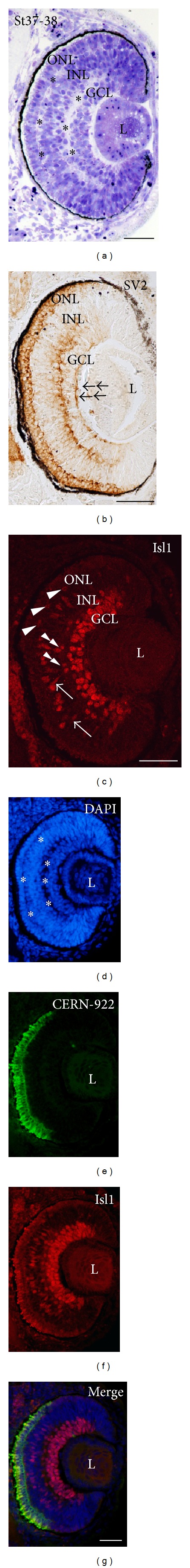
Figure 5. Morphological features and expression patterns of Isl1 and other cell differentiation markers in the St37/38 Xenopus laevis retina. (a) Toluidine blue-stained transverse retinal resin sections showed that the plexiform layers were observed across the central to mid-peripheral extent of the retina (asterisks). An immature GCL, 2-3 cells in depth, was also observed at this stage. (b) SV2 immunoproducts were observed in the OFL (double arrows), IPL, and ONL. (c) Abundant nuclei were immunoreactive for Isl1 in the GCL, but also in the INL. Thus, nuclei located in the amacrine cell layer (double arrowheads), bipolar cell layer (arrows), and horizontal cell layer (arrowheads) were detected with this antibody. (d)–(g) Incipient plexiform layers were also clearly distinguishable with the DAPI nucleic acid stain (asterisks in (d)). CERN-922 immunoreactivity paralleled the expression of Isl1 and extended towards the more mid-peripheral region of the retina. GCL: ganglion cell layer; INL: inner nuclear layer; L: lens; ONL: outer nuclear layer. Scale bars: 50 μm. Image published in: Álvarez-Hernán G et al. (2013) Copyright © 2013 Guadalupe Álvarez-Hernán et al. Creative Commons Attribution license Larger Image Printer Friendly View |