XB-IMG-118782
Xenbase Image ID: 118782
|
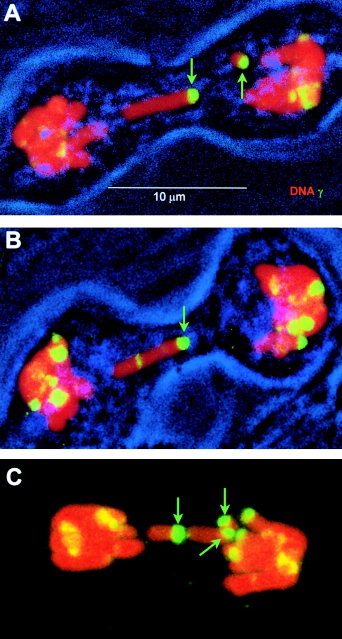
Figure 7. γ-H2AX foci on defective M. muntjak mitotic figures (maximum projections). Selected mitotic figures were imaged in M. muntjak cell cultures that had been exposed to 0.6 Gy on ice and covered with growth media at 37°C for 90 min before fixation. Green arrows point to ends of isolated chromosome arms with γ-H2AX foci. In A and B, transmitted light was collected to show the outline of the cell membrane. Image published in: Rogakou EP et al. (1999) © 1999 The Rockefeller University Press. Creative Commons Attribution-NonCommercial-ShareAlike license Larger Image Printer Friendly View |