XB-IMG-122779
Xenbase Image ID: 122779

|
Fig. 3
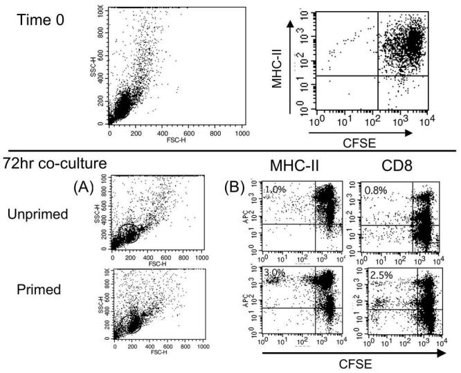
Detection of Xenopus primed splenocyte proliferation induced in vitro by FV3 infected FV3 peritoneal leukocytes using the CFSE assay. Splenocytes from an uninfected control frog or a frog primed 3 weeks before the assay by infection with FV3 were CFSE stained and co-cultured for 72 hours with FV3 infected peritoneal leukocytes obtained from the same frogs 3 days before the assay. Total culture was surface stained for MHC class II (AM20) or CD8 (AM22) and analyzed by FACS. A time zero analysis shows initial CFSE staining of splenocytes prior to co-culture. Analysis at 72 hrs (B) was done on gated population in the side scatter dot plot (A). Numbers in upper left quadrant indicates the percent of CD8 or Class II positive cells with diluted CFSE. Image published in: Morales H and Robert J (2008) Image downloaded from an Open Access article in PubMed Central. Article © by the author(s). This paper is Open Access and is published in Biological Procedures Online under license from the author(s). Copying, printing, redistribution and storage permitted. Journal © 1997-2008 Biological Procedures Online. Larger Image Printer Friendly View |
