XB-IMG-124564
Xenbase Image ID: 124564
|
|
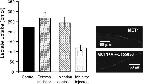
Figure 4. Microinjection of AR-C155858 inhibits MCT1 expressed in Xenopus oocytesMCT1 was expressed in Xenopus oocytes for 72 h prior to inhibitor treatment and assay of L-[14C]lactate uptake over 2.5 min. For addition of AR-C155858 internally, 20 oocytes were individually injected with 9.2 nl of 1 mM AR-C155858 or DMSO as a control, incubated for 5 min in 5 ml of pH 6 transport buffer and washed once prior to transport assay. For incubation with the equivalent amount of AR-C155858 added externally, 20 aliquots (9.2 nl) of 1 mM AR-C155858 were added to 5 ml of pH 6 transport buffer (final concentration of 35 nM) and incubated with the oocytes for 5 min prior to a single wash and transport assay as above. Uptake was corrected for the uptake by water-injected eggs under the same conditions and are presented as means±S.E.M. of 18–20 separate oocytes. The two images on the right-hand side show the plasma membrane expression of MCT1 in control oocytes and those incubated for 1 h with 1 μM AR-C155858 revealed by immunofluorescence microscopy. Image published in: Ovens MJ et al. (2010) © 2010 The Author(s) The author(s) has paid for this article to be freely available under the terms of the Creative Commons Attribution Non-Commercial Licence (http://creativecommons. Creative Commons Attribution-NonCommercial license Larger Image Printer Friendly View |