XB-IMG-170294
Xenbase Image ID: 170294
|
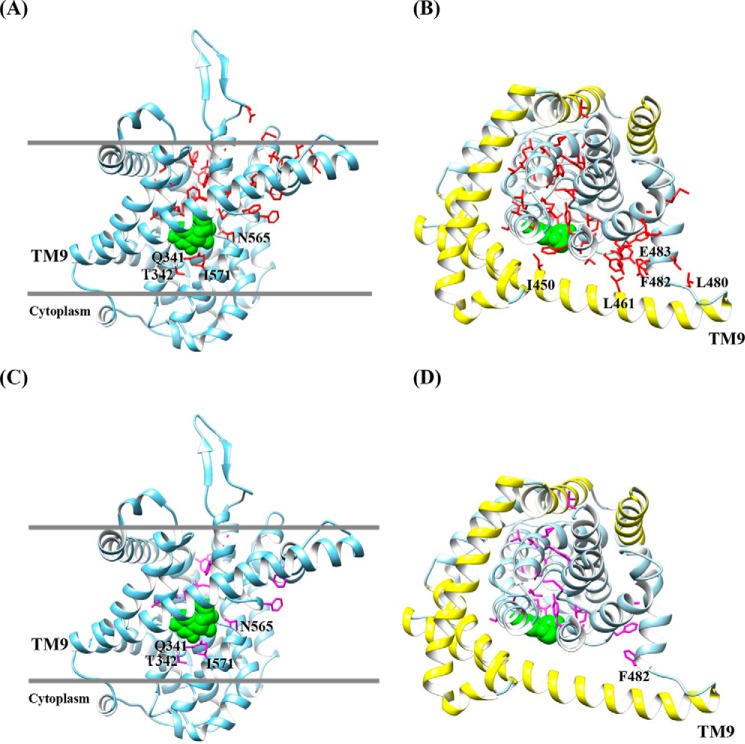
Figure 10. Homology model of hCNT3. Shown are cartoon representations of hCNT3 3D models based upon the crystal structure of the bacterial nucleoside transporter vcCNT (Protein Data Bank accession code 3TIJ) using the program SWISS-MODEL (56). Molecular graphics and analyses were performed with the UCSF Chimera package (57). A and C, models of hCNT3 viewed parallel to the membrane. The extracellular boundaries of the hydrophobic core of the bilayer predicted using the PPM server are shown as gray lines (58). B and D, models of hCNT3 viewed from the extracellular surface of the membrane. The outer scaffold domain of hCNT3 (TM4, TM5, IH1, TM6, and TM9) is shown in yellow. For clarity the loop linking HP2 and TM10 is not shown. Side chains of PCMBS-sensitive residues (A and B) are shown in red. Side chains of PCMBS-sensitive and uridine-protected residues (C and D) are shown in purple. The bound uridine molecule is shown in space filling representation (green). Image published in: Mulinta R et al. (2017) © 2017 by The American Society for Biochemistry and Molecular Biology, Inc. Creative Commons Attribution license Larger Image Printer Friendly View |