XB-IMG-124611
Xenbase Image ID: 124611

|
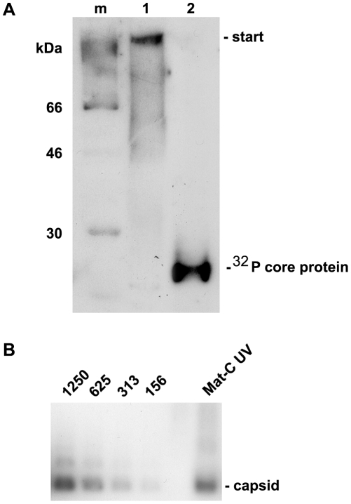
Figure 5. Analysis of cross-linked core particles.A. Lane 1: 32P-labelled Mat-C-UV and (lane 2) 32P-labelled Mat-C were separated on a 4–12% SDS-PAGE. Phosphoimaging showed that the core proteins of Mat-C-UV did not enter the separating gel indicating successful cross-linking. In contrast the core proteins of Mat-C migrated as a 21.5 kDa band. m: 14C-labelled molecular weight marker. B. Immune blot of Mat-C-UV and non cross-linked P-rC after native agarose gel electrophoresis. Mat-C-UV migrated as the P-rC standard indicating that the core proteins were linked within the individual capsids and that the capsids were not linked to each other. The identical migration indicates that UV irradiation has not changed the surface charge. The numbers on top of the standard dilution series give the amount of the P-rC in pg. Image published in: Schmitz A et al. (2010) Schmitz et al. Creative Commons Attribution license Larger Image Printer Friendly View |
