XB-IMG-126416
Xenbase Image ID: 126416
|
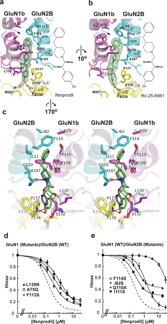
Figure 3. Phenylethanolamine binding sitea-b, Bindings of ifenprodil (a) and Ro 25-6981 (b) take place at the GluN1b-GluN2B subunit interface. Mesh represents Fo-Fc omit electron density map contoured at 3σ. Residues with asterisks in panel a are the ones previously shown to affect ifenprodil sensitivities. Adjacent to the binding pocket is an empty space surrounded by hydrophobic residues including GluN1b Ala75, GluN2B Ile82 and Phe114 (arrows). c, Comparison of binding patterns of ifenprodil (gray) and Ro 25-6981 (lime) in stereoview. The Ro 25-6981-bound structure is coloured as in panel b whereas the ifenprodil-bound structure is coloured white. d, New residues found to interact with phenylethanolamines in this study are mutated and analyzed for sensitivity to ifenprodil. Mutation of the residues surrounding the binding site caused changes in IC50 as well as extent of inhibition. Image published in: Karakas E et al. (2011) Image downloaded from an Open Access article in PubMed Central. Image reproduced on Xenbase with permission of the publisher and the copyright holder. Larger Image Printer Friendly View |