XB-IMG-127322
Xenbase Image ID: 127322

|
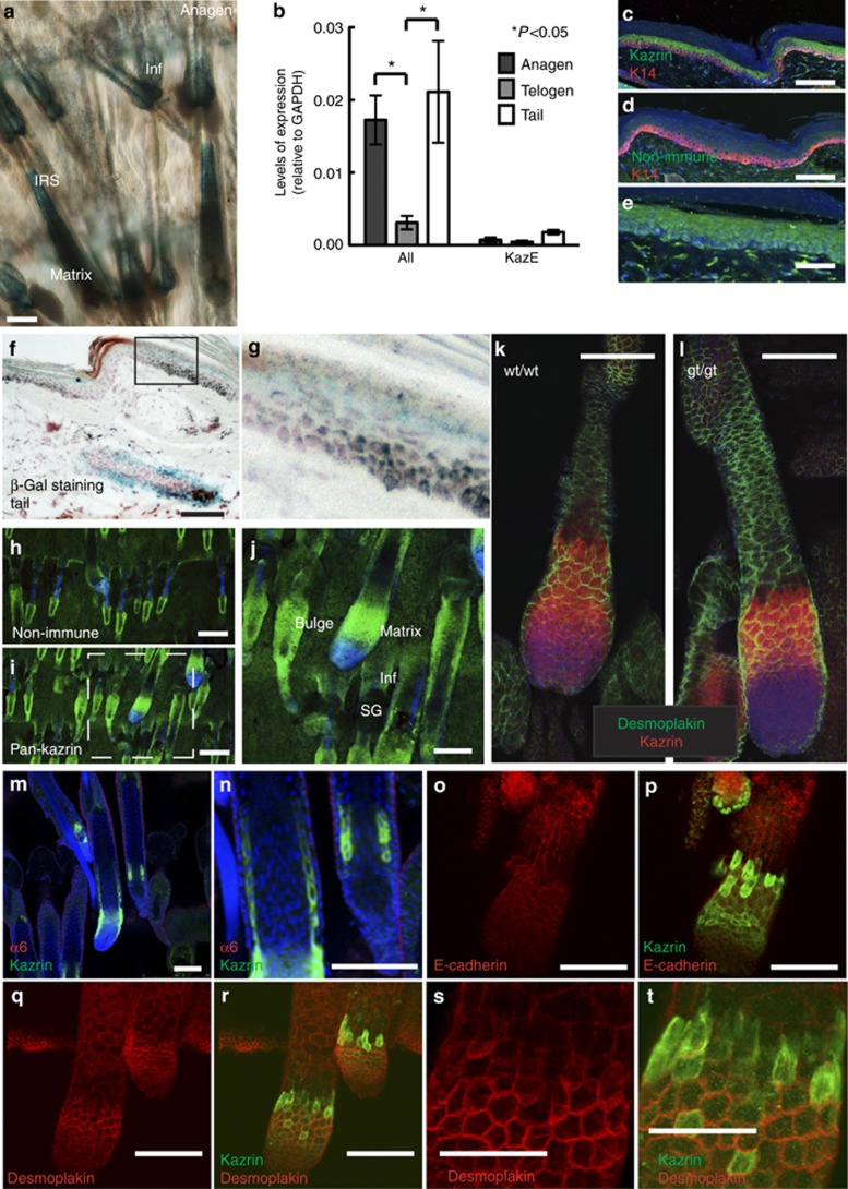
Figure 6. Kazrin expression in adult mice. (a) Epidermal tail whole mount of kazrin gene-trap (gt/gt) mouse stained for X-gal to visualize endogenous kazrin in hair during anagen. Inf, infundibulum; IRS, inner root sheath. (b) Quantitative RT-PCR of mRNA from anagen back skin, telogen back skin, or telogen tail skin using primers to detect all forms of kazrin (exons 6 and 7) or specific for kazrinE (KazE) (exons 10 and 11). The unpaired Student's t-test was used to determine whether differences in transcript levels were statistically significant. (c–e) Paraffin sections of tail-scale interfollicular epidermis from kazrin gt/gt mouse co-stained with antibodies to (c, d; red) K14 and (c, e; green) kazrin or (d; green) non-immune serum with (blue) 4′,6-diamidino-2-phenylindole (DAPI) nuclear counterstain. Field shown in (e) partially overlaps with the field shown in c. (f, g) Frozen section of tail skin from kazrin gt/gt mice stained for (blue) X-gal and counterstained (with nuclear fast red). (g) Higher-magnification view of the area demarcated by the black box in f. (h–t) Tail epidermal whole mounts stained with (h) non-immune serum, or the antibodies indicated. (j) Higher-magnification view of the area demarcated with white dashed line in i. (c–e, h–n) Blue staining is the DAPI nuclear counterstain. (j–t) The epidermis from wt/wt mice, except (l), which is from gt/gt mouse. Bars: (a, c, d, f, j–r) 100 μm; (e) 40 μm; (h, i) 200 μm; and (s, t) 50 μm. Image published in: Chhatriwala MK et al. (2012) Copyright © 2012 The Society for Investigative Dermatology, Inc. Creative Commons Attribution-NonCommercial-NoDerivatives license Larger Image Printer Friendly View |
