XB-IMG-127122
Xenbase Image ID: 127122
|
|
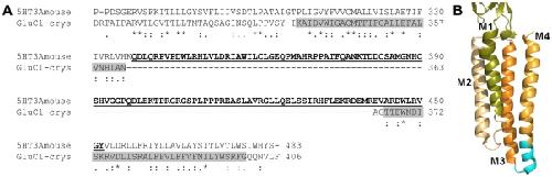
Figure 6. Sequence alignment GluClcrys and 5-HT3A and cartoon illustrating transmembrane region of GluClcrys.(A) Sequence alignment of the C-terminal region of mouse 5-HT3A receptor and GluClcrys. The GluClcrys M3 and M4 α helical membrane-spanning segments are indicated by the gray rectangles. The M3M4 loop region replaced by two alanines in the 5-HT3A-A2 construct is shown underlined in bold. The 5-HT3A-A2 construct yielded functional receptors with channel kinetics similar to wild type 5-HT3A receptors. Note that the 5-HT3A-A2 construct lacks 16 residues aligned with the cytoplasmic end of the GluCl M4 segment. (B) Cartoon representation of the transmembrane domain of one subunit of GluCl (PDB: 3RHW). The cyan region at the cytoplasmic end of the M4 segment indicates the residues that were replaced by two alanine residues in the5-HT3A-A2 construct. Image published in: McKinnon NK et al. (2012) McKinnon et al. Creative Commons Attribution license Larger Image Printer Friendly View |