XB-IMG-122498
Xenbase Image ID: 122498
|
|
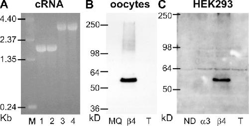
Figure 4. . cRNA gel-electrophoresis (A) and Western blots of expressed proteins in oocytes (B) and HEK293 cells (C). Approximately 1 μg of α3 (1), β4 (2), α3_β4 tandem (3), and β4_α3 tandem (4) besides the RNA ladder (M) were separated on a 1.5% agarose-gel (A). The Western blot in B was obtained from oocytes injected with water only (MQ), β4 only, or β4_α3 tandem (T) and the Western blot in C from HEK293 cells transfected with no DNA (ND), α3 only, β4 only, or β4_α3 tandem (T). Detection by β4 antibody and visualization by chemoluminescence. Bands for the β4 subunit were detected at the expected size of 56 kD for both blots after 30-s exposure. No breakdown products were observed for the tandem constructs in either blots, even for longer exposures up to 1 h. Note that the tandem fusion protein (predicted size of 115 kD) was not detected by the β4 antibody used (see text). Image published in: Groot-Kormelink PJ et al. (2004) Copyright © 2004, The Rockefeller University Press. Creative Commons Attribution-NonCommercial-ShareAlike license Larger Image Printer Friendly View |