XB-IMG-126721
Xenbase Image ID: 126721
|
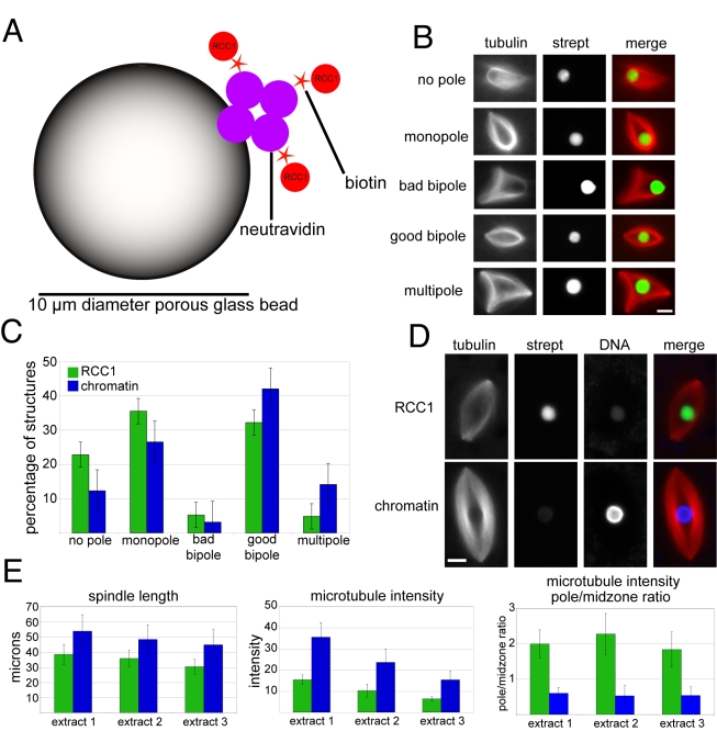
Figure 1. RCC1-coated beads induce spindle formation in Xenopus laevis egg extracts.(A) Schematic of a porous glass NeutrAvidin bead to which N-terminally biotinylated RCC1 is coupled, drawing not to scale. (B) Categories of microtubule structures formed around RCC1 beads. (C) Distribution of spindle structure categories formed around single RCC1 or chromatin beads. Nâ=â3 extracts, 80â130 structures counted per extract. (D) Images comparing representative RCC1 and chromatin bead spindles. Weak signals in the DNA channel of RCC1 bead spindle and the Alexa Fluor 488-labeled Streptavidin (strept) channel of chromatin bead spindle are due to slight bead autofluorescence. (E) Quantification of differences between RCC1 and chromatin bead spindle lengths, overall spindle microtubule intensity, and pole/midzone ratio of spindle microtubule intensity. Intensity is in arbitrary units. 80â130 structures counted per extract. Microtubules are red, RCC1 beads are green, and DNA beads are blue. Scale bars, 10 µm. Image published in: Halpin D et al. (2011) Halpin et al. Creative Commons Attribution license Larger Image Printer Friendly View |