XB-IMG-125168
Xenbase Image ID: 125168
|
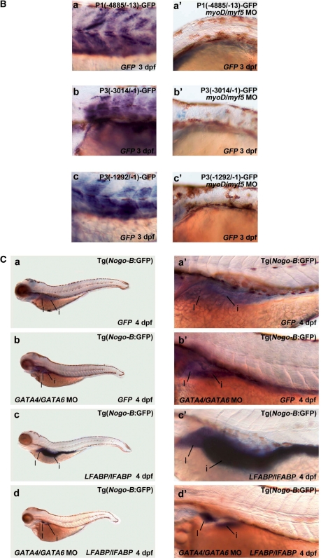
Figure 7. Loss of MyoD and Myf5 ablates somatic fast muscle and knockdown of GATA4 and GATA6 results in loss of GFP signal in the liver and intestine. (A) P1(−4885/−13)-GFP, P3(−3014/−1)-GFP and P3(−1292/−1)-GFP were separately injected or each coinjected with myod/myf5 double MOs into zebrafish embryos at the one-cell stage. Alternatively, myod/myf5 double MOs were injected to the transgenic Tg(Nogo-B:GFP) line at the one-cell stage. Zebrafish embryos at 48 hpf with GFP signals were selected for image analysis. Merged bright-field and fluorescence images are shown in panels (a’–h’), while fluorescence images are shown in panels (a–h). Scale bars indicate 100 μm. (B) Those embryos mentioned above at 3 dpf were subjected to whole-mount in situ hybridization using GFP as probe. All myod/myf5 double morphants did not show GFP signal. (C) GATA4/GATA6 double MOs were injected into zebrafish embryos of transgenic Tg(Nogo-B:GFP) line at one- to two-cell stage. The GATA4/GATA6 double-morphants and the parental transgenic line at 4 dpf were subjected to whole-mount in situ hybridization using GFP (panels a and b) and LFABP/iFABP (panels c and d) as probe. The liver and intestine were enlarged in panels (a’–d’). Image published in: Chen YC et al. (2010) © The Author(s) 2010. Creative Commons Attribution-NonCommercial license Larger Image Printer Friendly View |