XB-IMG-169064
Xenbase Image ID: 169064

|
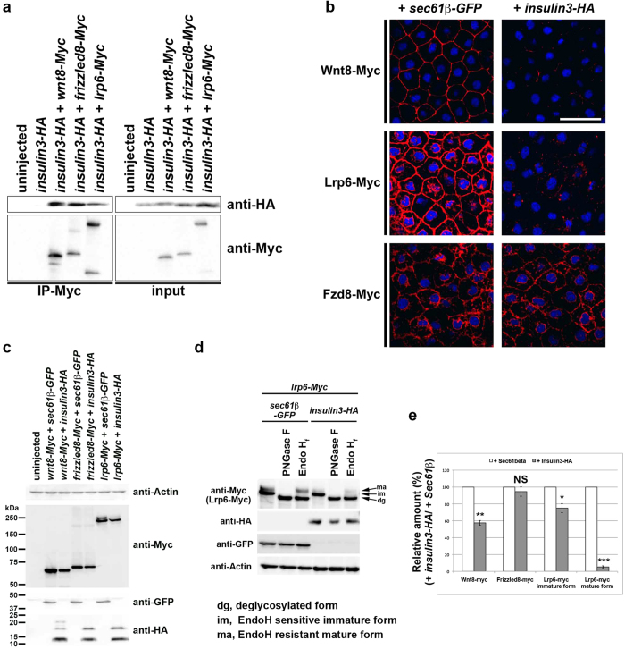
Figure 8. Insulin3 reduced the total amount of extra-cellular Wnt8 and membrane-localised Lrp6, but not Frizzled8.(a) Anti-Myc immunoprecipitation (IP). mRNAs was injected into the animal pole of both blastomeres at the two-cell stage in X. laevis embryos. One ng of insulin3-HA, wnt8-Myc, frizzled8-Myc, or lrp6-Myc mRNA was injected per embryo. Embryos were harvested at stage 10.5 for co-immunoprecipitation assay. (b,c,d) mRNAs were injected into the animal pole of both blastomeres at the two-cell stage in X. laevis embryos. Amounts of mRNA injected per embryos were: wnt8-Myc (500 pg), lrp6-Myc (500 pg), frizzled8-Myc (500 pg), sec61β-GFP (1 ng), and insulin3-HA (1 ng). Sec61β-GFP was used as an ER-localised control instead of cytoplasmic GFP because insulin3-HA was localised in ER (Fig. 7). (b) Confocal micrographs of ectodermal explants. Embryos were fixed and immunostained with anti-Myc antibody (red) for Wnt8-Myc, Lrp6-Myc, and Frizzled8-Myc at stage 11. Ectodermal explants were dissected after staining and mounted with DAPI. Insulin3 decreased cell-surface levels of Wnt8-Myc, Lrp6-Myc, but not Frizzled8-Myc. (c) Western blot analysis of embryo lysates. Insulin3 decreased the total amount of Wnt8-Myc. Insulin3 also reduced the mature form of Lrp6-Myc (upper band) which localised at the plasma membrane. Frizzled8-Myc had no effects. (d) Western blot analysis of embryo lysates treated with PNGaseF or EndoH as indicated. Insulin3 reduced EndoH-resistant mature form of Lrp6. dg, deglycosylated form; im, EndoH-sensitive immature form; ma, EndoH-resistant mature form. (e) Quantification of the Western blotting in (c) and (d). The protein bands in the blots from three independent experiments were quantified by using ImageJ for densitometry. The amount of protein in control (+Sec61β) was designated as 100%. Error bar indicate the SEM of these experiments. The data are presented as mean ± s.e.m. NS (not significant, P > 0.05), *P < 0.05; **P < 0.01; ***P < 0.001; (t-test, two tailed). Full-length blots are presented in Supplementary figure S9. Larger Image Printer Friendly View |
