XB-IMG-127551
Xenbase Image ID: 127551
|
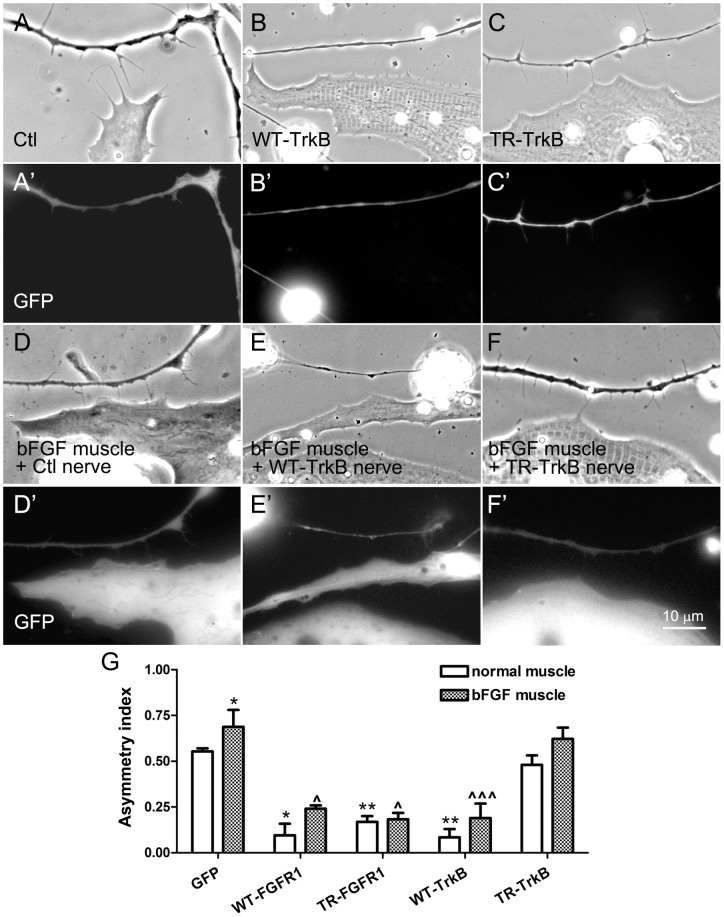
Figure 6. The influence of TrkB signaling on filopodial extension by neurons towards muscle.Nerve-muscle cocultures were prepared using neurons expressing GFP (A and A'), WT-TrkB (B and B') or TR-TrkB (C and C'). WT-TrkB-neurons, unlike control neurons, displayed little bias in the extension of filopodia towards muscle, but the TR-TrkB-neurons, which grew more filopodia than GFP-neurons, were able to send out filopodia preferentially in the direction of muscle. (D–F') On muscle cells expressing GFP plus bFGF, neurons expressing GFP (D and D') or TrkB proteins (E–F') were seeded. GFP-neurons extended even more filopodia towards muscle cells overexpressing bFGF (D and D') than towards normal muscle cells (A and A'). WT-TrkB-neurons once again grew fewer filopodia than GFP-neurons, but the bFGF-overexpressing muscle cells induced more filopodia in WT-TrkB-neurons (E and E') than control muscle cells (above). TR-TrkB-neurons also extended more filopodia towards bFGF-expressing muscle cells (F and F') than towards control cells. (G) Calculation of AI values for these cocultures as well as for those cultures in which neurons expressed FGFR1 proteins (pictures not shown). Asymmetric distribution of filopodia was slightly improved in neurons expressing WT-TrkB, TR-TrkB and WT-FGFR1, but not TR-FGFR1. Mean and SEM shown; t test: *p<0.05 and **p<0.01, compared to cocultures between Ctl neurons and normal muscle cells; ?p<0.05 and ???p<0.001, compared to cocultures made between Ctl neurons and bFGF-overexpressing muscle cells. Image published in: Li PP et al. (2012) Image reproduced on Xenbase with permission of the publisher and the copyright holder. Creative Commons Attribution license Larger Image Printer Friendly View |