XB-IMG-123483
Xenbase Image ID: 123483
|
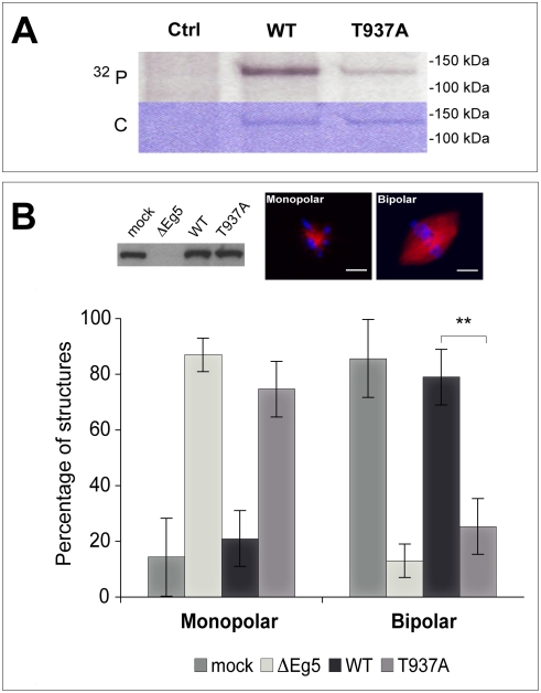
Figure 3. Spindles do not assemble in Eg5 depleted Xenopus egg extract in the presence of Eg5T937A.(A) Phosphorylation of wild-type Eg5 and of Eg5T937A in mitotic Xenopus egg extract, showing a strong reduction of the degree of phosphorylation of Eg5T937A. As a control, an extract without recombinant Eg5 is shown (Ctrl). Autoradiography (32P) and Coomassie-stained polyacrylamide gel (C) are shown. (B) Western blot (top left) showing the amount of Eg5 in mock depleted extract (mock), in Eg5 depleted extract (ΔEg5) and in Eg5 depleted extract after addition of Eg5 wild-type (WT) or of Eg5T937A (T937A). Fluorescence images of a monopolar spindle after Eg5 depletion (top middle) and of a bipolar spindle after ‘mock’ depletion (top right). Graphs (bottom) representing the percentages of monopolar and bipolar spindles under the conditions as indicated. More than 120 structures per condition were included in the analysis. ** indicates statistical difference (p = 0.0031, significance level 0.05). Spindle assembly cannot be rescued by Eg5T937A at the concentration used. Microtubules are TAMRA-labeled (red) and DNA is stained with Hoechst (blue). Scale bar is 10 µm. Image published in: Cahu J et al. (2008) Cahu et al. Creative Commons Attribution license Larger Image Printer Friendly View |