XB-IMG-175647
Xenbase Image ID: 175647
|
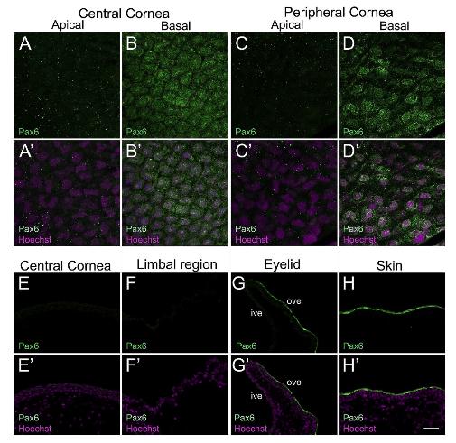
Fig. 3. Confocal and fluorescence light microscopic images showing immunofluorescent staining for Paired box protein 6 (Pax6) in larval and adult frog epithelia. (AâD) Pax6 (green) localizes to the basal epithelial cells of central and peripheral cornea in the tadpoles, as labeled. (Aâ²-Dâ²) Merged images for A-D with Hoechst labeled nuclei (magenta). (A-Aâ²) Pax6 expression is undetectable in apical cells of the central cornea. (B-Bâ²) Both the nucleus and cytoplasm of basal cells in the central corneal epithelium show uniform Pax6 staining. However, expression is excluded from the peri-nuclear region in these cells. Pax6-positive cells have a tight packing arrangement. (C-Câ²) Pax6 is not expressed in the apical cells of peripheral corneal epithelium. (D-Dâ²) Basal cells in the peripheral cornea also express Pax6. Fewer Pax6-positive cells were detected in the peripheral region. (EâH) Cross-sections of adult frog cornea, eyelid and skin stained with Pax6 (green) antibody, as labeled. The apical surface is located towards the top of each image and the basal surface towards the bottom. (Eâ²-Hâ²) Merged images for E-H with Hoechst labeled nuclei (magenta). (E-Eâ²) Pax6 staining is not observed in the central cornea. (F-Fâ²) Pax6 expression is also undetected in the limbal region. (G-Gâ²) Pax6 staining is noted only in apical cells of the outer surface of the ventral eyelid, where expression is primarily localized to the cytoplasm of these cells. (H-Hâ²) A similar pattern of Pax6 expression is seen in apical cells in the skin epithelium, outside the cornea. ive, inner ventral eyelid; ove, outer ventral eyelid. Scale bar in Hâ² equals 25â¯Î¼m for A-D, Aâ²-Dâ², and 50â¯Î¼m for E-H, Eâ²-Hâ. (For interpretation of the references to colour in this figure legend, the reader is referred to the Web version of this article.) Image published in: Sonam S et al. (2019) Copyright © 2019. Image reproduced with permission of the Publisher, Elsevier B. V.
Image source: Published Larger Image Printer Friendly View |