XB-IMG-124265
Xenbase Image ID: 124265

|
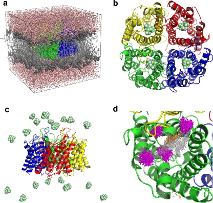
Fig. 1. a Simulation system of an hAQP1 tetramer (green, gray, red, and yellow space-filling representation) embedded in a POPE lipid bilayer (gray) and surrounded by water (red, white). b Starting positions for TEA_dockMD as described in the text. The tetramer is represented as cartoons and TEA in space-filling representation. c Starting positions for TEA_20random. Twenty TEA molecules were randomly placed in the bulk water. d TEA binding site defined via the distribution of TEA nitrogen positions obtained from TEA_dockMD (gray spheres), TEA_20random (magenta spheres), and umbrella sampling (orange spheres) Image published in: Müller EM et al. (2008) Image downloaded from an Open Access article in PubMed Central. © Springer-Verlag 2007 Larger Image Printer Friendly View |
