XB-IMG-125349
Xenbase Image ID: 125349
|
|
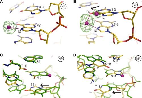
Figure 3. Co2+/Ni2+-DNA binding site selectivity. (A and B) Ni2+ (A) and Co2+ (B) binding to GG dinucleotide sites at locations 61 and −35/−34, respectively. Displacement of the 3′ guanine into the major groove at position −34 (arrow) allows N7/O6 cross-linking and creates a secondary, low occupancy, site for N7 coordination to the 3′ base. An anomalous difference electron density map, contoured at 8σ (A) and 4σ (B), is shown superimposed on the model. (C and D) The guanine base atoms at locations 8 (C) or 14 (D) from the Ni2+-NCP structure (gold carbon atoms) were least-squares superimposed onto those of the Mn2+-NCP structure (green) to illustrate how conformational alterations associated DNA stretching and histone–DNA register shifting can influence metal binding. In both instances, Co2+/Ni2+ binding occurs in the stretched particle half, whereas Mn2+ association is not observed at the respective locations. This appears to be largely a consequence of steric clearance provided by positive slide at the TG elements (arrow) associated with the change in DNA orientation. (A–D) Co2+/Ni2+ ions and coordinate (-covalent) bonds appear as magenta spheres and lines, respectively. Image published in: Mohideen K et al. (2010) © The Author(s) 2010. Creative Commons Attribution-NonCommercial license Larger Image Printer Friendly View |