XB-IMG-117640
Xenbase Image ID: 117640
|
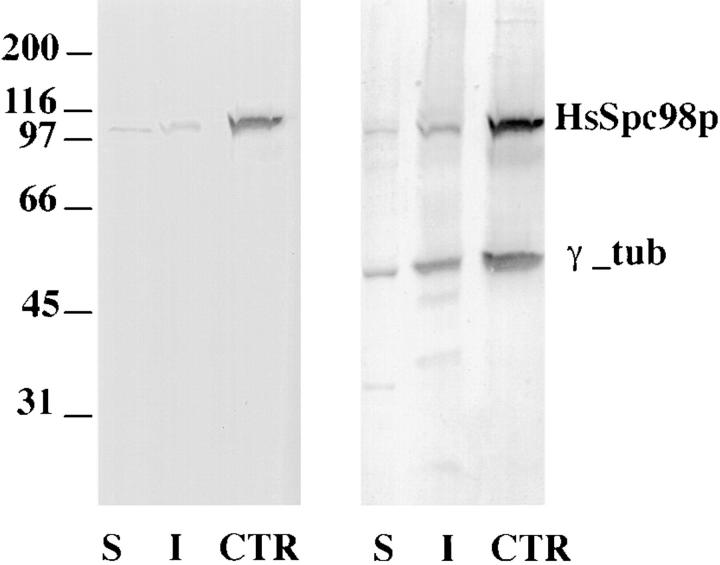
Figure 4. Western blot analysis of low-speed, Triton X-100–soluble (S) and -insoluble (I) protein fractions from unsynchronized KE37 cells and of a highly enriched centrosome preparation (CTR). Proteins were probed with affinity-purified HsSpc98p IgG (left). A band at 103 kD is observed in all fractions, while highly enriched in the centrosome fraction. The same blot was subsequently probed with anti–γ-tubulin (right). Note the similar partition of both proteins in all fractions. 10 μg of Triton X-100–soluble and -insoluble proteins representing 2 × 10⁵ and 6 × 10⁵ cells, respectively, and ∼3 × 10⁷ centrosomes were loaded. Image published in: Tassin AM et al. (1998) Image reproduced on Xenbase with permission of the publisher and the copyright holder. Creative Commons Attribution-NonCommercial-ShareAlike license Larger Image Printer Friendly View |