XB-IMG-126563
Xenbase Image ID: 126563
|
|
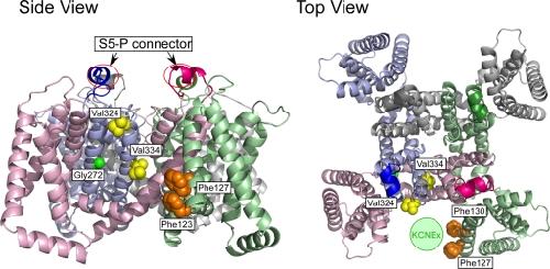
Figure 12. Locations of important amino acid residues determined in this study and a putative binding site of the KCNE protein. The structural model for the open state of human KCNQ1 (Smith et al., 2007) is used to locate Phe123 (hidden below Phe127 in the top view), Phe127 and Phe130 (hidden behind Phe127 in the side view) on the S1 segment (orange), Gly272 on the S5 segment (green), and Val324 and Val334 on the S6 segment (yellow). S5-P connectors are circled in the side view (left). All four α subunits are shown, and each subunit is in a different color. The hypothetical location for the KCNE protein (“KCNEx”) is indicated as a light green circle in the top view (right). Image published in: Nakajo K et al. (2011) © 2011 Nakajo et al. Creative Commons Attribution-NonCommercial-ShareAlike license Larger Image Printer Friendly View |