XB-IMG-121979
Xenbase Image ID: 121979
|
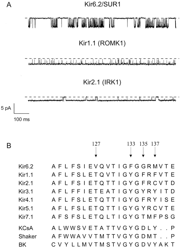
Figure 2. (A) Single-channel currents recorded at −60 mV from an inside-out patch excised from an oocyte expressing Kir6.2/SUR1, Kir1.1, or Kir2.1, as indicated. Currents were recorded in a symmetrical 140-mM solution. The dashed line indicates the zero current level. (B) Sequence of the pore loop of Kir6.2 compared with that of some other types of K+ channel. Arrows indicate the mutated residues. Image published in: Proks P et al. (2001) © 2001 The Rockefeller University Press. Creative Commons Attribution-NonCommercial-ShareAlike license Larger Image Printer Friendly View |