XB-IMG-127051
Xenbase Image ID: 127051
|
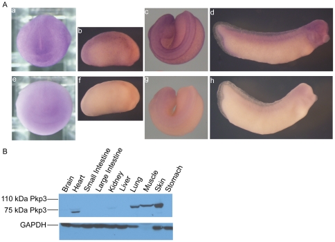
Figure 3. Xenopus Pkp3 spatial expression profiles.(A) Whole-mount in situ RNA hybridization reveals Pkp3 mRNA signals in the anterior and dorsal neural fold regions of neurula embryos (subpanel a). At elongation stages (stage 22; subpanel b), staining of the skin remains apparent, as does a concentration in dorsal structures. At tadpole stages, neural derived tissues including the brain, branchial arches and the spinal cord are stained, as are somites (subpanels câd). As a basis for comparison (negative controls), we undertook sense-probe hybridizations in parallel (subpanels EâH). (B) Immuno-blotting on adult Xenopus tissue extracts. We used an affinity purified rabbit polyclonal antibody directed against the N-terminal domain of Pkp3, which clearly resolved the Pkp3 protein isoform migrating at approximately 75 kDa. This product was strongly expressed in heart, lung, muscle and skin. Weak, reproducible expression was detected in brain and kidney. Image published in: Munoz WA et al. (2012) Munoz et al. Creative Commons Attribution license Larger Image Printer Friendly View |