XB-IMG-123575
Xenbase Image ID: 123575
|
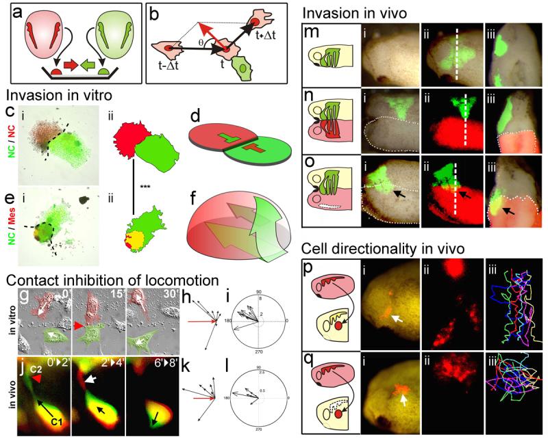
Figure 2. Contact Inhibition of Locomotion in NC cells in vitro and in vivo(a) Experimental design. (b) Analysis of Contact Inhibition of Locomotion. Mean velocities were measured Δt minutes before and after the collision. Acceleration (red) was calculated for each cell. Angle of collision calculated after initial trajectory alignment (θ). (c-f) Invasion of confronted explants. (c) There is no invasion in NC/NC confrontations (i), outlines in (ii), overlapping area in yellow; schematised in (d). (e) NC explants completely invade and cover mesodermal explants (i), outlines in (ii), overlapping area in yellow; schematised in (f). Green arrows in f: NC path of invasion (see Supplementary Fig. 3 for supporting confocal images). (g-l) Contact Inhibition of Locomotion. (g) Collision between two pseudocoloured NC cells in vitro. Time in minutes. White arrows: direction of migration; red arrowhead: collision. (h) Velocity vectors for NC in vitro, initial velocity vector (red arrow). (i) Acceleration vectors for NC collisions in vitro. They are clustered after the collision (p<0.005, n=10). (j) Collision of two NC cells (C1, C2) in vivo shown as the difference between two consecutive two-minute frames. Green: new area; red: collapsing area; black arrow: direction of migration; red arrowhead: cell contact; white arrow: collapsing protrusion. (k) In vivo velocity vectors. (l) In vivo acceleration. They are clustered after the collision (p<0.01, n=10). (m-o) NC invasion in vivo. i, ii: lateral view; iii: transverse section along the dashed line showed in ii. NC cells are not able to invade an adjacent embryo that has NC (n; 0% of invasion, n=15), but they can to invade an embryo without NC (o; arrow, 80% of invasion, n=10). (p, q) Cell directionality in vivo. A small group of Nuclear-RFP-labelled NC cells were grafted into a normal embryo (p) or in an embryo in which the NC were previously removed (q). Note that grafted cells migrate directionally in the intact embryo (persistence: 0.6±0.04, n= 30), but not when the host NC were removed (persistence: 0.2±0.02, n=20). Image published in: Carmona-Fontaine C et al. (2008) Image downloaded from an Open Access article in PubMed Central. Image reproduced on Xenbase with permission of the publisher and the copyright holder. Larger Image Printer Friendly View |