XB-IMG-118964
Xenbase Image ID: 118964
|
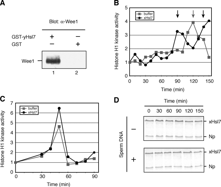
Figure 2. Excess xHsl7 accelerates entry into mitosis in a nuclear-dependent manner. (A) GST-yeast Hsl7 (lane1) or GST (lane 2) were incubated in Xenopus egg extracts, retrieved on glutathione Sepharose, and immunoblotted with anti-Wee1 antibodies. (B) xHsl7-encoding mRNA or buffer was incubated in cycling extracts with sperm nuclei (5,000/μl) and an ATP-regenerating system. Aliquots were stored at the indicated times and assayed for their ability to phosphorylate histone H1 in the presence of [32P]ATP. Phosphorylated histone was resolved by SDS-PAGE, subjected to autoradiography, and quantified by phosphoimager with the zero time point normalized to 1. Square, buffer; circle, FLAG-xHsl7 mRNA addition. Arrows indicate the time of the nuclear envelope break down and the chromosome condensation as monitored by microscope. (C) Same assay as panel B except that sperm nuclei were absent. (D) In vitro–translated 35S radiolabeled xHsl7 and nucleoplasmin (as an internal control) were added to cycling extracts with or without nuclei present. Samples were withdrawn at the indicated times, resolved by SDS-PAGE, and subjected to autoradiography to monitor xHsl7 stability. Mitosis was observed at 90 min by fluorescence microscopy of extracts containing Hoechst-stained nuclei. Image published in: Yamada A et al. (2004) Copyright © 2004, The Rockefeller University Press. Creative Commons Attribution-NonCommercial-ShareAlike license Larger Image Printer Friendly View |