XB-IMG-117028
Xenbase Image ID: 117028
|
|
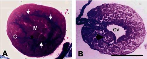
Figure 7. Histological transverse cross-section (8 μm) of presumptive male (A) and female (B) leopard frog (R. pipiens) at metamorphosis (Gosner stage 46). Gonads are not completely differentiated. Note the intact cortex (C) and medulla (M) separated by blue connective tissue (arrows in A). Also note medullary regression and ovarian vesicle (OV) but absence of significant oogenesis in the female (B). A single oocyte (arrow) is noted in the female. Scale bar = 125 μm. Image published in: Hayes TB et al. (2006) Image downloaded from an Open Access article in PubMed Central. This is an Open Access article: verbatim copying and redistribution of this article are permitted in all media for any purpose, provided this notice is preserved along with the article's original DOI Larger Image Printer Friendly View |