XB-IMG-125469
Xenbase Image ID: 125469
|
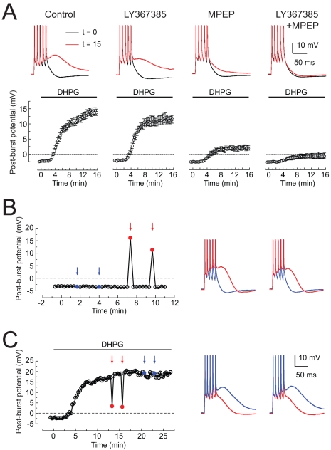
Figure 2. Metabotropic glutamate receptors mediating the DHPG-induced ADP.(A) Top: sample responses to bursts of five action potentials in normal ACSF (black) or following application of 2.5 µM DHPG (red) in four different conditions. Drug concentrations are 25 µM LY367385 and 10 µM MPEP. Bottom: time course of medium ADP amplitude (58 ms after last spike) following application of 2–4 µM DHPG beginning at t = 0 min. (B) Post-burst potential plotted over time in normal ACSF. On select trials, 500 µM DHPG was applied locally, via pressure application from a broken patch pipette (see Materials and Methods) approximately 3 s before the current-injection stimulus. Blue arrows and traces indicate application to the distal apical dendrites (near the border of stratum radiatum and stratum lacunosum-moleculare); red arrows and traces indicate application to the perisomatic region (stratum pyramidale). (C) Same as (B) except 2–4 µM DHPG was applied to the bath beginning at t = 0 min and normal ACSF was applied using localized pressure application to the perisomatic (red) or apical dendritic (blue) regions. In all panels, the stimulus is five brief current injections and the interval between trials is 20 s. Action potentials are truncated. Image published in: Park JY et al. (2010) Park et al. Creative Commons Attribution license Larger Image Printer Friendly View |